




Abstract
This review provides a comprehensive overview of lipid-based nanoparticle synthesis methods, spanning traditional and top-down approaches used for liposomes, solid lipid nanoparticles (SLNs) and nanostructured lipid carriers (NLCs) to advanced bottom-up nanoprecipitation technologies for modern lipid nanoparticle (LNP) formulation. It provides deeper insight into the most widely adopted and advanced methods — such as microfluidics — for achieving controlled, efficient, and reproducible LNP formulation, with precise control over particle size, PDI, encapsulation efficiency, and morphology to meet the stringent demands of drug development.
What are the main methods for lipid-based nanoparticle formulation?
Numerous methods are available for the synthesis of lipid-based nanoparticles, a broad family that includes liposomes, solid lipid nanoparticles (SLNs), nanostructured lipid carriers (NLCs), and modern lipid nanoparticles (LNP) designed for nucleic acid delivery. Throughout this review, “lipid-based nanoparticles” refers to this broader family of systems, whereas “LNPs” specifically refers to modern lipid nanoparticles developed for nucleic acid delivery.
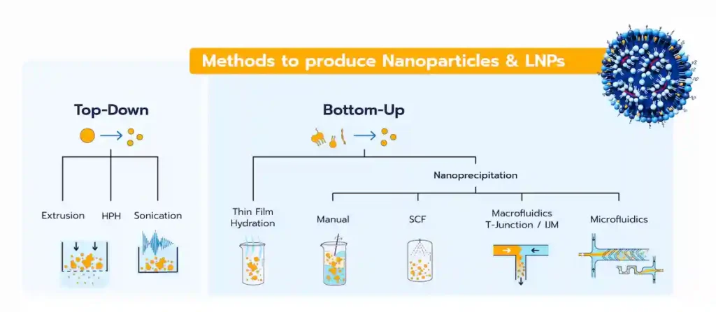
Lipid-based nanoparticle formulation methods can broadly be categorized into two approaches based on how the nanoparticles are formed (Figure 1):
- Top-down methods: Also referred to as high-energy methods, these involve applying energy to break larger particles into smaller ones.
- Bottom-up methods: These rely on the self-assembly of lipid components to form nanoparticle systems.
Modern RNA-LNP formulation predominantly relies on bottom-up approaches, particularly nanoprecipitation, which can be performed using different mixing technologies ranging from manual mixing to macrofluidic and microfluidic systems.
This review examines these formulation approaches, from traditional preparation techniques to modern controlled mixing technologies, highlighting their principles, advantages, limitations, and suitability across different stages of lipid-based nanoparticle development.
How does the formulation method influence LNP quality?
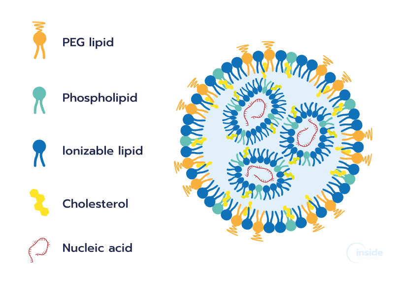
The development of successful LNPs depends on multiple factors, including the selection of appropriate lipids, their ratios, and concentrations. However, the formulation process itself is equally important, as the method and associated process parameters directly influence the critical quality attributes (CQAs) of the resulting nanoparticles, including particle size, polydispersity index (PDI), encapsulation efficiency, and morphology (Figure 2). These attributes can, in turn, affect LNP stability and biological performance, including biodistribution, cellular uptake, therapeutic efficacy, and safety.
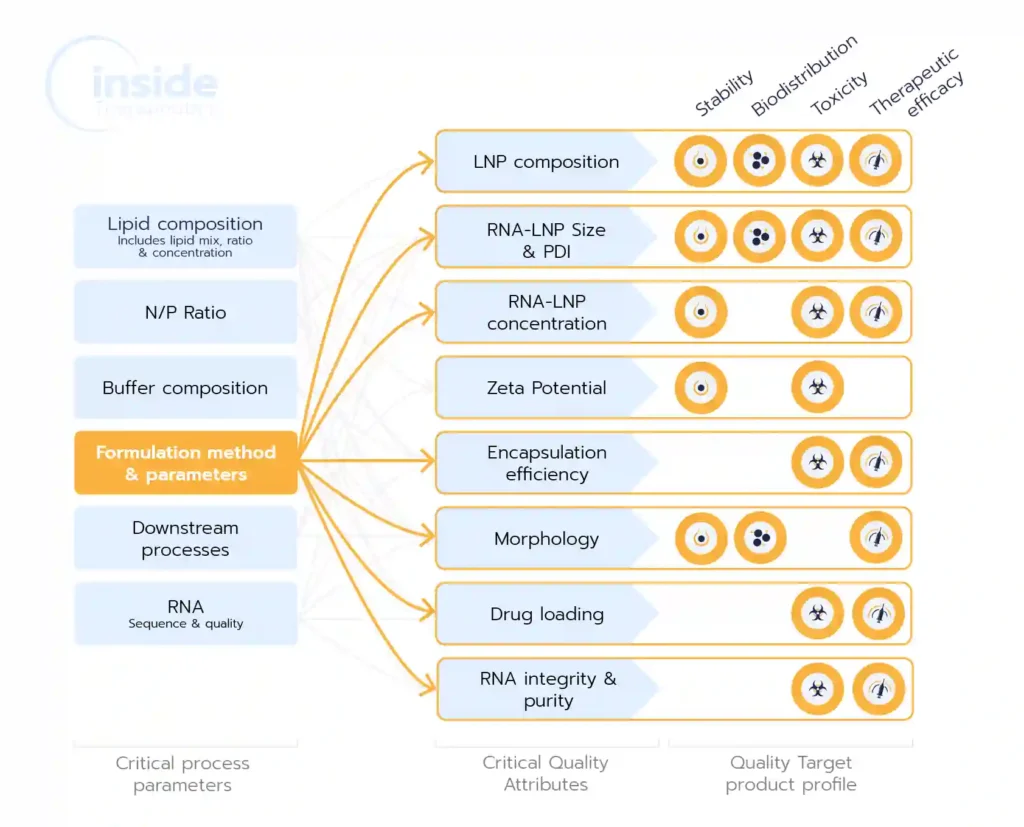
Given this relationship between formulation conditions, LNP properties, and biological performance, selecting an appropriate formulation method is an important step in LNP development. The choice should reflect the requirements of each development stage, including available material, required batch volume, process control and reproducibility, and the ability to increase production scale while maintaining the desired LNP quality.
Establishing a well-controlled and reproducible formulation process early in development can therefore facilitate progression toward larger-scale production while reducing the need for extensive process re-optimization. Maintaining consistent process conditions and LNP quality across scales remains an important challenge during LNP formulation scale-up and, ultimately, the translation of formulations toward clinical manufacturing.
How do LNP formulation requirements evolve from screening to manufacturing?
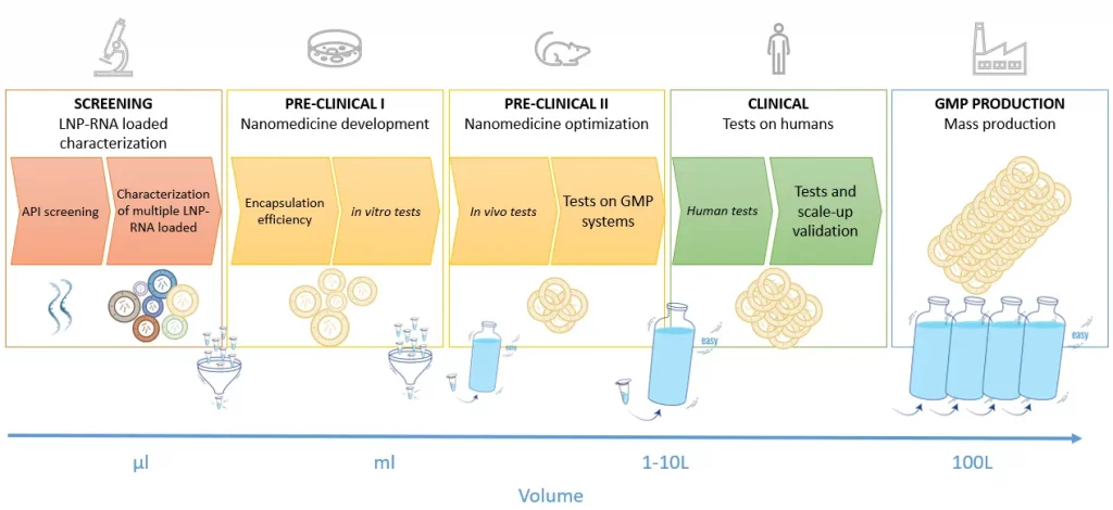
The requirements for LNP formulation evolve considerably throughout the drug development process. Early-stage development prioritizes low-volume, high-throughput screening, whereas later stages require larger batch volumes, greater process robustness, and ultimately manufacturing under appropriate quality standards (Figure 3).
During early discovery and formulation screening, many combinations of RNA payloads, lipid compositions, molar ratios, concentrations, and process conditions may need to be evaluated to identify promising formulations. This applies across applications such as mRNA vaccines and gene-editing approaches. Because RNA and specialized lipids can be costly and available in limited quantities, screening methods should ideally operate at low volumes while providing sufficient control and reproducibility to compare formulations reliably. High-throughput capabilities can further accelerate the evaluation of large formulation libraries.
As candidates progress through development, the number of formulations typically decreases while the required batch volume increases. Selected formulations move from initial in vitro screening toward in vivo studies and, eventually, clinical development and larger-scale production. At this stage, reproducibility and process scalability become increasingly important. Ideally, the transition between scales should minimize changes in the conditions governing LNP formation, helping to maintain consistent CQAs and biological performance as production volume increases. This process continuity represents an important consideration during LNP scale-up.
As development progresses toward clinical trials and commercial production, additional requirements related to process robustness, quality control, reproducibility, and good manufacturing practice (GMP)-compliant manufacturing become increasingly important. The formulation technology selected during development should therefore be considered not only in terms of its immediate experimental requirements, but also in terms of its ability to support progression toward larger and more controlled production scales.
What are top-down methods for lipid-based nanoparticle synthesis and what are their limitations?
Top-down methods apply energy to reduce larger lipid structures, droplets, or particles into smaller and more homogeneous nanoparticle populations. Depending on the technique, size reduction can result from shear forces, turbulence, cavitation, or passage through membranes with defined pore sizes. Representative approaches include high-pressure homogenization (HPH), ultrasonication, and membrane extrusion.
These methods have been widely used for the preparation of lipid-based nanoparticles, particularly liposomes, solid lipid nanoparticles (SLNs), and nanostructured lipid carriers (NLCs). They may be used directly during nanoparticle preparation or as a secondary size-reduction step following another formulation method. For example, thin-film hydration is commonly followed by membrane extrusion to reduce particle size and polydispersity in liposome preparation.
Top-down technologies are well established, and some can be readily implemented at large scale. However, their suitability depends strongly on the nanoparticle system and payload. High mechanical energy, repeated processing, and — in some methods — elevated temperatures can be undesirable for sensitive biological components.
High-pressure homogenization (HPH)
High-pressure homogenization (HPH) is a well-established technique for the preparation of lipid-based nanoparticles, particularly solid lipid nanoparticles (SLNs) and nanostructured lipid carriers (NLCs). During HPH, a lipid-containing dispersion (molten lipids, water, and surfactants) is forced through small orifices under high pressure. The resulting high shear forces, turbulence, and cavitation disrupt larger lipid structures and reduce them to the nanoscale range. [1]
One of the main advantages of HPH is its short processing time and scalability, making it suitable for both laboratory-scale preparation and larger-scale production. However, the process is energy-intensive, and the mechanical energy generated during homogenization can increase the temperature of the formulation, which may be problematic for thermosensitive compounds.
For SLNs and NLCs, HPH is commonly performed using two approaches: hot homogenization and cold homogenization. In both cases, the active compound is initially incorporated into the lipid phase, while the subsequent processing conditions differ in how the lipid dispersion is formed and homogenized.
Cold homogenization HPH
In cold homogenization, the active compound is first incorporated into a molten lipid phase, which is then rapidly cooled and solidified. The resulting solid lipid is reduced to microparticles, dispersed in a cooled aqueous surfactant solution, and homogenized at or below room temperature to form nanoparticles. This approach limits thermal exposure during homogenization, making it suitable for temperature-sensitive compounds, although it generally produces larger particles than hot homogenization. [1]
Hot homogenization HPH
In hot homogenization, the active compound is incorporated into a molten lipid phase, which is dispersed in a heated aqueous surfactant solution to form a pre-emulsion. The pre-emulsion is then homogenized above the lipid melting temperature and subsequently cooled, causing the lipid phase to solidify and form nanoparticles. This approach is efficient, but the elevated processing temperatures may be unsuitable for thermosensitive compounds. [1]
Ultrasonication
Ultrasonication is a dispersion-based method typically associated with the preparation of SLNs and NLCs. The active compound is incorporated into a molten lipid phase and dispersed in a heated aqueous surfactant solution to form an emulsion. The emulsion is then exposed to high-frequency ultrasound, generating cavitation and localized shear forces that reduce droplet size. Upon cooling, the lipid phase solidifies to form a nanoparticle dispersion.
The main advantage of this method is its simplicity and accessibility, as it can be performed using standard laboratory equipment. However, sonication may lead to broad particle size distributions, while prolonged probe sonication introduces a risk of metal contamination due to probe erosion. [1]
Ultrasonication can also be used in liposome preparation, particularly to reduce the size and polydispersity of pre-formed liposomes.
Membrane extrusion
Membrane extrusion reduces and homogenizes particle size by repeatedly passing a pre-formed lipid dispersion through membranes with defined pore sizes. It is commonly used following methods such as thin film hydration to generate smaller and more homogeneous nanoparticle populations.
Although extrusion provides relatively good control over particle size and size distribution, it requires pre-formed particles and additional processing steps, and its throughput is limited by the membrane area and operating conditions. It is therefore primarily useful for laboratory-scale preparation and post-formation size control, particularly in liposome preparation. [1]
How do bottom-up approaches enable LNP formation through self-assembly?
Unlike top-down methods, bottom-up approaches form nanoparticles through the assembly of molecular components rather than the size reduction of pre-existing particles. Lipids or other building blocks organize into nanoscale structures as a result of changes in their physicochemical environment, such as solvent composition, concentration, pH, or mixing conditions.
Several preparation techniques can be considered bottom-up approaches, including thin-film hydration and nanoprecipitation-based methods. For modern RNA-LNP formulation, nanoprecipitation driven by rapid mixing of an organic lipid phase with an aqueous RNA phase has become particularly important.
Thin film hydration
The thin film hydration method, also known as the Bangham method, is a classical technique widely used for liposome preparation. The process involves dissolving lipids in an organic solvent, creating a thin lipid film via solvent evaporation, and subsequently hydrating the film with an aqueous solution to trigger the spontaneous formation of lipid vesicles.
Thin-film hydration is accessible but generally provides limited control over particle size and size distribution, often producing relatively heterogeneous and multilamellar vesicle populations. For this reason, it is frequently followed by additional size reduction or homogenization steps, such as membrane extrusion or sonication. [2]
Although historically important for liposome preparation, thin-film hydration is not the predominant approach used for modern ionizable RNA-LNP formulation.
How does nanoprecipitation drive RNA-LNP self-assembly?
Nanoprecipitation is the primary bottom-up approach used for modern RNA-LNP formulations. In a typical process, an organic phase containing the lipid components — commonly dissolved in ethanol — is rapidly mixed with an aqueous phase containing the nucleic acid payload.
As the two phases mix, rapid dilution of the organic solvent changes the local solvent environment and decreases lipid solubility. This drives lipid nanoprecipitation and self-assembly into nanoparticles. For RNA-LNPs containing ionizable lipids, formulation under acidic conditions also promotes protonation of the ionizable lipid and electrostatic interactions with negatively charged RNA, contributing to efficient payload encapsulation. Rapid solvent exchange is now a standard route for RNA-LNP assembly.

Nanoprecipitation triggered self-assembly process involves solvent exchange, lipid supersaturation, nucleation, growth, and subsequent particle stabilization. The relative rates of these processes strongly influence the physicochemical characteristics of the resulting LNPs. As introduced in our lipid nanoparticle formation review, LNP properties are therefore highly dependent on formulation composition and process conditions. Important parameters include:
- Mixing rate: Faster and more homogeneous mixing generally promotes rapid solvent exchange and can result in smaller, more uniform LNPs by limiting particle growth and aggregation.
- Phase ratio: The relative proportions of the aqueous and organic phases influence the rate of ethanol dilution and lipid supersaturation, and this might affect particle size and dispersity.
- Lipid concentration: Increasing the concentration of formulation components can increase the frequency of molecular and particle interactions during self-assembly, often promoting particle growth and increasing LNP size.
- Lipid composition and stabilizing components: Lipid identity and concentration influence particle nucleation, growth, and stabilization. In particular, PEG-lipids can limit particle growth, with increasing PEG-lipid content generally associated with smaller LNPs.
- Buffer and solvent conditions: pH, ionic strength, and solvent composition influence lipid ionization, RNA–lipid interactions, and the kinetics of self-assembly, particularly for ionizable RNA-LNP formulations.
- Temperature and fluid properties: Temperature, viscosity, and related fluid properties can affect diffusion, solvent exchange, and mixing kinetics, thereby influencing nanoparticle formation.
Because nanoprecipitation commonly involves ethanol, the resulting LNP dispersion generally undergoes downstream processing to remove residual solvent and exchange the formulation buffer. Processes such as dialysis, centrifugal ultrafiltration, and tangential flow filtration (TFF) are commonly used for these purposes and can themselves influence final product quality and stability.
The fundamental nanoprecipitation principle remains similar across different formulation technologies; what primarily differs is how rapidly, uniformly, and reproducibly the aqueous and organic phases are mixed. This has led to the development of mixing approaches ranging from simple manual methods to controlled macrofluidic and microfluidic systems.
Manual & batch methods
Manual LNP mixing can induce nanoprecipitation through pipetting, vortexing, or other bulk-mixing approaches. Similar principles can be implemented at larger volumes using batch mixing vessels.
These methods are simple, inexpensive, and accessible, making them useful for preliminary proof-of-concept experiments. However, limited control over local mixing and solvent exchange results in greater variability in final nanoparticle characteristics and batch-to-batch reproducibility. The influence of mixing conditions on LNP formation and the differences between manual and controlled microfluidic mixing are discussed in more detail in our review on LNP hand mixing and microfluidic mixing.
For systematic formulation screening and development, more controlled mixing technologies are therefore generally preferred when precise and reproducible control over LNP quality is required.
Supercritical fluid methods (SCFs)
Supercritical fluid (SCF) technologies represent an alternative bottom-up approach for the preparation of lipid-based nanoparticles, with the potential to reduce the use of conventional organic solvents or facilitate their removal. Supercritical CO₂ is most commonly used because of its relatively accessible critical conditions and ease of removal after processing.
Several SCF-based processes have been investigated, including supercritical antisolvent (SAS) precipitation. In SAS, supercritical CO₂ interacts with an organic solution containing the formulation components and acts as an antisolvent, promoting precipitation as the solvent environment changes. [3]
Although SCF approaches can offer advantages in solvent removal and particle processing, they require specialized high-pressure equipment and careful control of phase behavior and component solubility. Their use remains limited compared with conventional nanoprecipitation-based approaches, particularly for modern RNA-LNP formulation.
Impingement jet mixing/T-junction mixing
Impingement jet mixers (IJMs) and T-mixers achieve rapid nanoprecipitation by bringing the organic lipid phase and aqueous phase together at high velocity. The resulting intensive mixing rapidly dilutes the organic solvent and generates the conditions required for lipid self-assembly and nanoparticle formation.
Their high throughput and compatibility with continuous processing make these systems particularly attractive for larger-scale LNP production. [2] However, effective mixing generally requires relatively high flow rates, resulting in larger minimum working volumes than those typically required during early-stage formulation screening. Particle formation can also be highly sensitive to flow conditions, while conventional IJM configurations generally operate within a relatively restricted flow rate ratio (FRR) close to 1:1.
These characteristics make IJM well suited to larger-scale production, but less flexible for low-volume screening and broad process-parameter exploration.
Microfluidic mixing
The need to precisely control the rapid solvent exchange governing nanoprecipitation has made microfluidic mixing a widely used approach for RNA-LNP formulation. Microfluidic systems manipulate small volumes of fluid within microscale channels, enabling precise control over flow rates, phase ratios, mixing conditions, and formulation volume.
At the microscale, fluid flow is commonly laminar. Because molecular diffusion alone can result in relatively slow mixing, microfluidic devices use engineered channel geometries to shorten diffusion distances or generate controlled chaotic advection, accelerating the mixing of the aqueous and organic phases.
For RNA-LNP formulation, two important process parameters are the total flow rate (TFR) and flow rate ratio (FRR). Together with lipid concentration, composition, buffer conditions, and mixer geometry, these parameters influence solvent exchange and nanoparticle self-assembly and can consequently affect nanoparticle characteristics such as particle size, polydispersity, encapsulation efficiency, and morphology. [2]
A practical implementation of microfluidic RNA-LNP formulation, including organic and aqueous phase preparation, selection of TFR and FRR, mixing, downstream processing, and characterization, is described in our RNA-LNP formulation protocol using TAMARA.
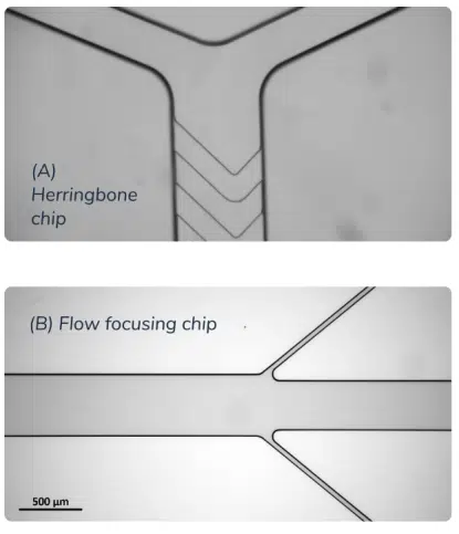
Several microfluidic mixer geometries have been developed to control the mixing process during RNA-LNP formulation (Figure 5), each with its own advantages and limitations. These include T- and Y-junction mixers, hydrodynamic flow-focusing systems, staggered herringbone micromixers, baffle mixers, and toroidal mixers, which use different mechanisms to accelerate mixing and therefore offer different operating ranges in terms of sample volume, flow rate, and throughput. For a detailed comparison of these microfluidic architectures, see our RNA-LNP formulation technologies review.
How to choose the right LNP formulation method?
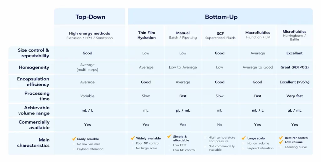
As discussed above, lipid-based nanoparticles encompass different systems that are not necessarily prepared using the same technologies. HPH is well established for SLNs and NLCs, while thin-film hydration and extrusion remain widely used for liposomes. For modern RNA-LNPs, formulation predominantly relies on controlled nanoprecipitation, implemented through mixing approaches ranging from manual mixing to microfluidic and macrofluidic technologies. As summarized in Table 1, each method presents different trade-offs in terms of sample volume, process control, reproducibility, throughput, and scalability. Consequently, the choice of formulation method is critically influenced by the stage of drug development.
During early-stage LNP screening and formulation optimization, minimizing material consumption while maintaining precise and reproducible control over nanoparticle formation is particularly important. Microfluidic mixing has emerged as the gold standard in this phase, as it enables controlled nanoprecipitation at low volumes and systematic exploration of formulation and process parameters. Microfluidic platforms such as TAMARA apply this principle to RNA-LNP formulation across screening and preclinical-scale volumes. However, the throughput of conventional microfluidic systems can become limiting as batch-volume requirements increase, and scale-up may require parallelization, alternative mixer geometries, or a transition to a different mixing technology.
At larger production scales, technologies capable of higher throughput or continuous processing become increasingly important. Impingement jet mixers and related macrofluidic approaches can support large-volume RNA-LNP production, while high pressure homogenization is well established for the large-scale preparation of SLNs, NLCs, and other lipid-based nanoparticles. Although IJMs and macrofluidic LNP formulation approaches are well-suited for meeting the demands of mass production, they may trade off some degree of precision compared to microfluidics and their minimum volume requirements limit their suitability for early-stage screening.
Ultimately, selecting the most appropriate LNP formulation method necessitates a comprehensive understanding of the intended application, the physical and chemical properties of the LNPs, and the regulatory and practical requirements of the production process. As outlined, a seamless transition between early-stage development and large-scale manufacturing remains a critical challenge. Addressing this gap will require novel technical innovation to enable a more streamlined and efficient LNP development pipeline. To explore this scale up challenge in more detail and how we are addressing it through NanoPulse, our novel mixing technology designed to maintain consistent formulation conditions across scales, read our review on RNA-LNP scale-up from screening to GMP manufacturing.
Table 1. Comparison of representative top-down and bottom-up methods for lipid-based nanoparticle formulation.
How do you characterize LNP quality after formulation?
As discussed earlier, the choice of the LNP formulation method and associated process parameters significantly influences critical quality attributes (CQAs) of the nanoparticles. This section explores the key CQAs and examines how formulation conditions affect these attributes.
Comprehensive characterization is essential throughout LNP development to verify that the formulation meets its intended quality profile and to assess batch-to-batch consistency. Characterization becomes increasingly important as formulations progress toward preclinical and clinical development, where reproducible control of product quality must be demonstrated.
While the regulatory landscape for nanomedicines continues to develop, existing characterization guidelines and established practices provide a framework for evaluating nanoparticle-based therapeutics. Among the most commonly evaluated LNP attributes are particle size and size distribution, zeta potential, morphology and internal structure, and encapsulation efficiency. Go deeper on the topic in our LNP and liposomes characterization review.
LNP size and size distribution

Particle size is one of the key physicochemical attributes of LNPs, as it can influence biodistribution, cellular uptake, and ultimately biological performance. Particle size is determined by the combined effects of lipid composition, component concentrations, mixing technology used during nanoparticle formation, and formulation conditions.
The optimal particle size therefore depends on the route of administration, target tissue, and intended application rather than on a single universal value. For RNA-LNP therapeutics, particle sizes are commonly within the 50–200 nm range.
It is worth noting that, counterintuitively, LNP size is mostly driven by lipid composition and formulation conditions rather than by the size of the encapsulated RNA cargo.
Particle size is commonly evaluated using techniques such as dynamic light scattering (DLS) and nanoparticle tracking analysis (NTA).
The polydispersity index (PDI) provides information about the width of the particle size distribution obtained from DLS measurements. PDI values range from approximately 0 to 1, with lower values indicating a narrower and more homogeneous particle population. In RNA-LNP development, PDI values below approximately 0.2 are generally indicative of a relatively narrow size distribution.
Consequently, formulation development often involves screening different lipid compositions alongside process conditions to identify combinations that provide the desired LNP characteristics. LNP Starter Kits can facilitate this process by enabling the comparison of clinically relevant lipid compositions during early-stage RNA-LNP screening, while the RNA-LNP Formulation Calculator can help translate selected compositions and formulation parameters into the required lipid and RNA quantities.
LNP zeta potential
Zeta potential represents the electrical potential at the slipping plane, the boundary between the particle and the surrounding fluid where the particle and its associated layer of ions move relative to the bulk solution, rather than the actual charge directly at the particle surface.
Zeta potential can influence colloidal interactions, protein adsorption, cellular interactions, and biodistribution, making it a useful parameter during LNP characterization. While a high absolute zeta potential can provide electrostatic stabilization in some colloidal systems, modern ionizable RNA-LNPs frequently exhibit relatively low or near-neutral surface potentials under physiological conditions and can remain stable through steric stabilization provided by PEG-lipids and other surface components.
Zeta potential is commonly determined using electrophoretic light scattering (ELS), in which nanoparticle electrophoretic mobility under an applied electric field is measured and used to estimate the zeta potential. These measurements should always be interpreted in the context of measurement pH, buffer composition, ionic strength, and formulation composition.
LNP morphology and internal structure
Modern RNA-LNPs do not necessarily adopt a single architecture, and multiple structural organizations have been reported depending on lipid composition, payload, and formulation conditions.
Cryogenic transmission electron microscopy (Cryo-TEM) is particularly valuable for direct visualization of LNPs in a near-native hydrated state, enabling assessment of particle morphology, internal organization, and population heterogeneity. Complementary scattering techniques such as small-angle X-ray scattering (SAXS) and small-angle neutron scattering (SANS) can provide population-level information about internal organization. These methods can help investigate features such as lipid organization, lamellarity, characteristic structural distances, and the spatial distribution of nanoparticle components.
Together, these techniques can reveal structural differences arising from the conditions under which LNPs are formed. In particular, the mixing environment during nanoprecipitation can influence lipid–RNA assembly and therefore the morphology and internal organization of the resulting particles. A recent head-to-head study comparing 11 mixing conditions identified distinct Cryo-TEM morphologies among RNA-LNPs produced using different mixing approaches, further demonstrating the relationship between mixing conditions and LNP structure. See how different mixing technologies affect RNA-LNP structure and performance. [4]
Encapsulation efficiency, encapsulation yield, and drug loading
Encapsulation efficiency (EE%), encapsulation yield (EY%), and drug loading provide complementary information about LNP formulation quality, process recovery, and payload-carrying capacity. Although these terms are sometimes used interchangeably, they describe different aspects of the formulation process.
Encapsulation efficiency (EE%) represents the proportion of the payload present in the final sample that is encapsulated within the nanoparticles rather than remaining free in solution. For RNA-LNPs, high EE% is desirable because it indicates effective association of the RNA payload with the nanoparticle fraction, while excessive free RNA can contribute to unwanted biological effects and compromise formulation performance. In academia, EE% is commonly determined using fluorescence-based assays such as RiboGreen, which quantify free RNA and then total RNA following nanoparticle disruption. For a practical methodology, see our protocol for measuring RNA-LNP encapsulation efficiency using the RiboGreen assay. Alternative analytical approaches are also being explored to simplify RNA payload quantification and reduce sample preparation. In a collaborative Inside Therapeutics–Marama Labs study, scatter-free absorbance spectroscopy using CloudSpec showed EE% measurements consistent with the RiboGreen assay while enabling RNA quantification without nanoparticle lysis. The complete formulation and characterization workflow is described in our application note on CloudSpec payload quantification.
Encapsulation yield (EY%) describes the amount of encapsulated payload recovered relative to the amount initially introduced into the formulation process. Unlike EE%, EY% therefore captures material losses occurring during formulation and, depending on where it is measured, downstream processing. This distinction is particularly important when working with expensive or limited RNA payloads: a formulation can show high EE% while still exhibiting relatively poor overall RNA recovery.
Drug loading (DL) describes the amount of payload carried relative to the nanoparticle’s lipid content. It therefore provides information about the payload-carrying capacity of the formulation and is relevant to dosing and formulation concentration.
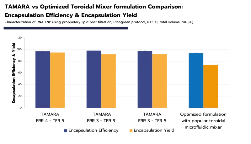
Encapsulation efficiency is highly dependent on the formulation method used. For RNA-LNPs, advanced techniques such as microfluidics can achieve encapsulation efficiencies exceeding 90% in optimized formulations.
However, EE% alone does not provide a complete picture of formulation performance. Evaluating EE%, EY%, and drug loading together provides a more informative assessment of both nanoparticle quality and process efficiency. This distinction is particularly relevant when comparing formulation technologies (Figure 8). Two systems may produce LNPs with similarly high EE% while differing substantially in RNA recovery, dead volume, or other process-related losses. Carefully selecting not only the formulation method but also the synthesis equipment itself therefore appears critical. For experimental data comparing encapsulation efficiency and encapsulation yield between TAMARA and a toroidal mixing platform, see our comparative RNA-LNP formulation study.
LNP formulation: Key takeaways and future perspectives
Lipid nanoparticles (LNPs) have become a leading non-viral delivery system for nucleic acid therapeutics, including mRNA, siRNA, saRNA, and pDNA. By protecting these payloads and facilitating their intracellular delivery, LNPs have enabled applications ranging from vaccines to gene therapy.
As discussed throughout this review, LNP quality depends not only on lipid composition and formulation design, but also on how the nanoparticles are produced. The formulation method and associated process parameters influence CQAs such as particle size, PDI, internal structure, and encapsulation efficiency, which can ultimately affect the biological performance of the resulting formulation. Selecting and controlling an appropriate formulation process is therefore an integral part of RNA-LNP development.
No single preparation method is optimal for every development stage. Within this space, microfluidic mixing provides precise and reproducible control at low volumes, making it particularly well suited to formulation screening and optimization. This approach underlies our platform TAMARA, which uses microfluidic mixing for RNA-LNP preparation from low-volume screening through preclinical-scale production. As volume requirements increase, however, throughput and process scalability become increasingly important, requiring technologies capable of extending this level of process control toward larger-scale production.
One of the key challenges in RNA-LNP development is maintaining consistent formulation conditions and LNP quality as processes move from early-stage screening to manufacturing. Advances in mixer design, continuous processing, process monitoring, and scalable formulation technologies are needed to address this gap and support the translation of complex RNA-LNP therapeutics. This process continuity challenge is one we are currently addressing with NanoPulse, a new mixing technology designed to extend controlled RNA-LNP formulation across scales.
References
[1] Musielak, E., Feliczak-Guzik, A., & Nowak, I. (2022). Synthesis and Potential Applications of Lipid Nanoparticles in Medicine. Materials, 15(2), 682. https://doi.org/10.3390/MA15020682
[2] Mehta M, Bui TA, Yang X, Aksoy Y, Goldys EM, Deng W. Lipid-Based Nanoparticles for Drug/Gene Delivery: An Overview of the Production Techniques and Difficulties Encountered in Their Industrial Development. ACS Materials Au. American Chemical Society; 2023. p. 600–19. https://doi.org/10.1021/acsmaterialsau.3c00032
[3] C. S. Kalpage and R. A. T. Isuranga, “Supercritical Fluid Technologies for Nanoparticle Production,” pp. 215–246, 2025, doi: 10.1007/978-981-96-9176-0_11.
[4] T. Bethiana et al., “Identifying differential effects from eleven mixing techniques on mRNA lipid nanoparticle physicochemistry and biological performance,” Nov. 09, 2025. doi: 10.1101/2025.11.07.687311.
Looking to get started with RNA-LNP formulation?
Reach out to us to learn how we can help!
Frequently asked questions on RNA-LNP formulation
What are the main methods used for LNP synthesis?
Lipid-based nanoparticle synthesis methods fall into two families. Top-down (high-energy) methods such as high-pressure homogenization and ultrasonication break bulk material down into nanoparticles and are commonly used for systems such as SLNs and NLCs. Bottom-up approaches rely on the self-assembly of lipid components into nanoparticles and include thin-film hydration and nanoprecipitation-based methods (e.g., impingement jet mixing and microfluidics). For modern RNA-LNP formulation, controlled nanoprecipitation using microfluidic or macrofluidic mixing technologies has become particularly important because it enables reproducible control over nanoparticle formation and payload encapsulation. Microfluidic mixing is today’s reference method for RNA-LNP formulation at the screening and preclinical R&D stage.
What is the difference between top-down and bottom-up lipid-based nanoparticle synthesis?
Top-down methods start from bulk lipid material and apply energy (pressure, shear, ultrasound) to reduce it to nanoparticle size. Bottom-up methods instead form nanoparticles through the self-assembly of individual lipid components. In RNA-LNP nanoprecipitation, this typically involves rapidly mixing a lipid-containing organic phase with an aqueous phase containing the RNA payload, triggering changes in lipid solubility, protonation, and lipid–RNA association that drive nanoparticle formation. Microfluidics in particular, based on nanoprecipitation triggered self-assembly, enables the production of high-quality LNPs with precise and reproducible control over their final characteristics, which is why it dominates modern RNA-LNP development at the screening and preclinical stage.
Why is microfluidics considered the gold standard for RNA-LNP formulation in early-stage development?
At the micrometer scale, fluids flow in a controlled, laminar regime, so the mixing that triggers LNP self-assembly can be tuned precisely through flow parameters (TFR and FRR). This translates into tight size control, narrow size distributions (PDI < 0.2), high encapsulation efficiency (> 80-90%), and strong batch-to-batch reproducibility — while working at the very low volumes that make early screening affordable. That combination is why microfluidics is the reference method for the early R&D stages of RNA-LNP formulation.
What is the difference between encapsulation efficiency (EE%) and encapsulation yield (EY%)?
They measure two different things and should be read together. Encapsulation efficiency (EE%) is the fraction of RNA encapsulated relative to the total RNA in the final sample — a high EE% means little free RNA remains, which limits toxicity. Encapsulation yield (EY%) – also known as RNA recovery – is the fraction of your starting RNA you actually recover encapsulated, so it reflects the real process losses (dead volumes, head-and-tail waste) of the equipment itself. A system can show excellent EE% while still wasting a large share of costly RNA in yield — which is why the choice of formulation system, not just the method, matters.
What parameters should you consider when choosing an LNP formulation method?
The appropriate LNP formulation method depends on the development stage, required formulation volume, payload, and desired level of process control. Important considerations include the required volume (microliters for screening, up to liters for production); the level of control & repeatability you need over the critical quality attributes such as size, PDI, encapsulation efficiency, morphology — since these drive therapeutic efficacy; the encapsulation yield and material losses, which weigh heavily when RNA and lipids are expensive; and batch-to-batch reproducibility. Two factors are easy to overlook: the ability to keep the same nanoparticle as you move up in scale, so early-stage results remain relevant at large-scale production, and practical process constraints such as material compatibility, residual-solvent requirements, and later GMP requirements.
Can I use the same LNP formulation method from screening to large-scale production?
Maintaining the same formulation process across the full development pathway remains challenging with the established mixing technologies. Microfluidics excels at low-volume screening but faces clogging and throughput limits when scaled up, while impingement jet mixing and related macroscale methods handle large volumes but cannot access the small volumes needed for screening. Switching method between scales changes the mixing — and therefore the nanoparticle itself — which is the core scale-up challenge in RNA-LNP manufacturing. Emerging technologies aim to close this gap by keeping the mixing identical across scales: NanoPulse (Inside Therapeutics), for example, relies on high-frequency alternating injection of two liquid phases to produce the same nanoparticle from microliters to liters.