




Abstract
Lipid nanoparticles (LNPs) are cutting-edge nanodrug delivery systems designed to protect and efficiently deliver nucleic acids, playing a pivotal role in mRNA vaccine success. This guide provides a complete overview of LNP formulation, explaining how lipid nanoparticles work and the factors that define their quality. It examines LNP composition — ionizable lipids, phospholipids, cholesterol, and PEG-lipids — detailing how each influences stability, encapsulation efficiency, and biodistribution. The review also compares key formulation methods, including microfluidics, for producing uniform, stable LNPs with high encapsulation rates.
What are lipid nanoparticles and how were they developed?
Resulting from 50 years of research in the nanocarrier delivery field, lipid nanoparticles (LNPs) are close cousins to other lipid-based nanoparticles (LBNPs), including liposomes and solid lipid nanoparticles (SLNs). Unlike these earlier generations, designed primarily to encapsulate hydrophobic and/or hydrophilic small molecules, LNPs have the unique capability of encapsulating nucleic acids — such as messenger RNA (mRNA) and small interfering RNA (siRNA) — and delivering them to a specific location in the body.
Composed of a lipid shell surrounding an internal core that entraps oligonucleotides, these non-viral delivery systems protect their nucleic acid cargo from enzymatic degradation while promoting its delivery to target cells.
These unique features open up immense possibilities for researchers who wish to develop their ribonucleic acid-based therapeutics and deliver them to a specific location in the body. This groundbreaking technology was brought to global attention by BioNTech and Moderna through the COVID-19 mRNA-LNP vaccines, marking the first worldwide deployment of mRNA therapeutics at population scale.
Throughout this guide, we will cover:
- Why lipid nanoparticles are well suited for nucleic acid delivery
- Their composition and structure
- Available formulation methods and their trade-offs
- Key characterization techniques and quality attributes
- Intracellular delivery mechanisms
Why are lipid nanoparticles widely used for nucleic acid delivery?
While other lipid-based nanoparticles can be used as a carrier for a wide range of small molecules, their structure is less suited for the efficient encapsulation and intracellular delivery of negatively charged nucleic acids, such as mRNA and siRNA, for several reasons:
1/ Weak electrostatic interactions between neutral lipid systems and negatively charged oligonucleotides can limit nucleic acid complexation and encapsulation efficiency.
2/ In addition to encapsulation challenges, successful nucleic acid delivery requires nanoparticles capable of promoting intracellular release while minimizing toxicity.
From liposomes to LNPs
Lipid nanoparticles share similarities with conventional liposomes but differ through one major innovation: the incorporation of ionizable lipids.
One main limitation of the traditional liposomal carriers is the poor interaction with nucleic acids due to their neutral charge. For this reason, cationic lipids were used to enhance interactions with negatively charged RNA, leading to improved encapsulation efficiency. Although these formulations enhanced encapsulation efficiency, their persistent positive charge was often associated with increased cytotoxicity and immune activation. To tackle this issue, a new kind of lipid known as ionizable lipid was engineered.
Table 1. Comparison of liposomes and lipid nanoparticles (LNPs).

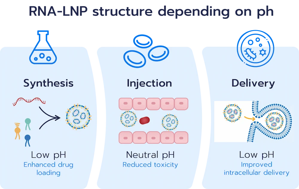
These ionizable lipids have the capability of switching charge with the pH, being positively charged at low pH while remaining neutral at physiological pH. This pH-dependent behavior enables efficient RNA encapsulation during nanoparticle formulation at low pH while improving biocompatibility following administration into the body.
In addition to better encapsulation, the use of the ionizable cationic lipids also play a critical role in promoting endosomal escape, a key step for efficient intracellular RNA delivery. Upon entering the endosome, where the pH value drops, ionizable lipids get protonated leading to particle destabilization and facilitating the release of the nucleic acid cargo into the cytoplasm of the cell.
The combination of ionizable lipids with helper lipids, cholesterol, and PEG-lipids enabled the development of a new generation of lipid-based nanoparticles with complex and formulation-dependent morphologies. Rather than exhibiting a single universal structure, LNPs are generally composed of an outer lipid organization surrounding internal lipid/RNA assemblies in which ionizable lipids interact with and stabilize nucleic acids.
By adjusting the lipid types and ratios, researchers can modulate key nanoparticle properties such as particle size, surface charge, encapsulation efficiency, stability, biodistribution, and intracellular delivery performance. As a result, optimizing LNP composition has become a major challenge in the development of efficient nucleic acid therapeutics.
Lipid nanoparticle composition
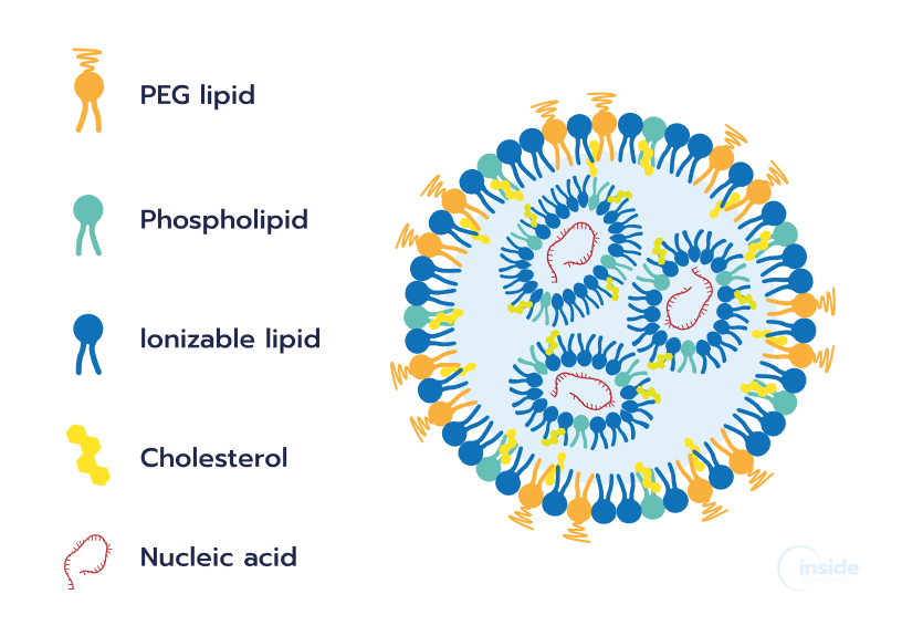
The lipid composition of LNPs plays a central role in determining their physicochemical properties, biological interactions, and therapeutic performance. Parameters such as particle size, encapsulation efficiency, stability, biodistribution, cellular uptake, and intracellular delivery are all strongly influenced by the nature and ratio of the different lipid components used during formulation.
Although LNP formulations can vary depending on the therapeutic application and nucleic acid payload, most RNA-LNP systems are generally composed of four main lipid constituents: an ionizable lipid (40 – 50 mol%), a helper phospholipid (10 – 15 mol%), cholesterol (38 – 50 mol%), and a polyethylene glycol (PEG)-lipid (1.5 – 2 mol%). Each component fulfills a distinct role in nanoparticle formation, stability, circulation behavior, and intracellular delivery, making lipid composition a critical parameter in the design and optimization of nucleic acid therapeutics.
Ionizable cationic lipid
These lipids remain predominantly neutral at physiological pH but become positively charged under acidic conditions, below their acid dissociation constant (pKa). This pH-responsive behavior enables several essential functions during RNA-LNP formulation and delivery.

1/ They promote electrostatic interactions with negatively charged nucleic acids, enabling efficient RNA encapsulation during nanoparticle formation.
2/ Following cellular uptake, their protonation within the acidic endosomal environment contributes to endosomal membrane destabilization, facilitating intracellular release of the nucleic acid cargo into the cytoplasm.
3/ At physiological pH, their predominantly neutral charge helps reduce systemic toxicity and immune activation compared with permanently cationic lipid systems.
Ionizable lipids generally represent the largest fraction of RNA-LNP formulations, typically accounting for approximately 40–50 mol% of the total lipid composition. Clinically validated ionizable lipids such as DLin-MC3-DMA (Alnylam Pharmaceuticals), SM-102 (Moderna), and ALC-0315 (Pfizer-BioNTech) have enabled the development of approved siRNA and mRNA-LNP therapeutics.

PEG-lipids
PEG-lipids typically represent only 1–2 mol% of an LNP formulation, yet they have a major influence on nanoparticle properties. Located at the particle surface, PEG chains create a hydrophilic steric barrier that reduces aggregation, improves colloidal stability, and promotes the formation of more homogeneous nanoparticle populations. PEG-lipids also influence particle size, biodistribution, circulation time, and immune recognition. However, excessive PEGylation may hinder cellular uptake and intracellular delivery, making the optimization of PEG content and lipid anchor structure an important aspect of LNP design.
Phospholipids
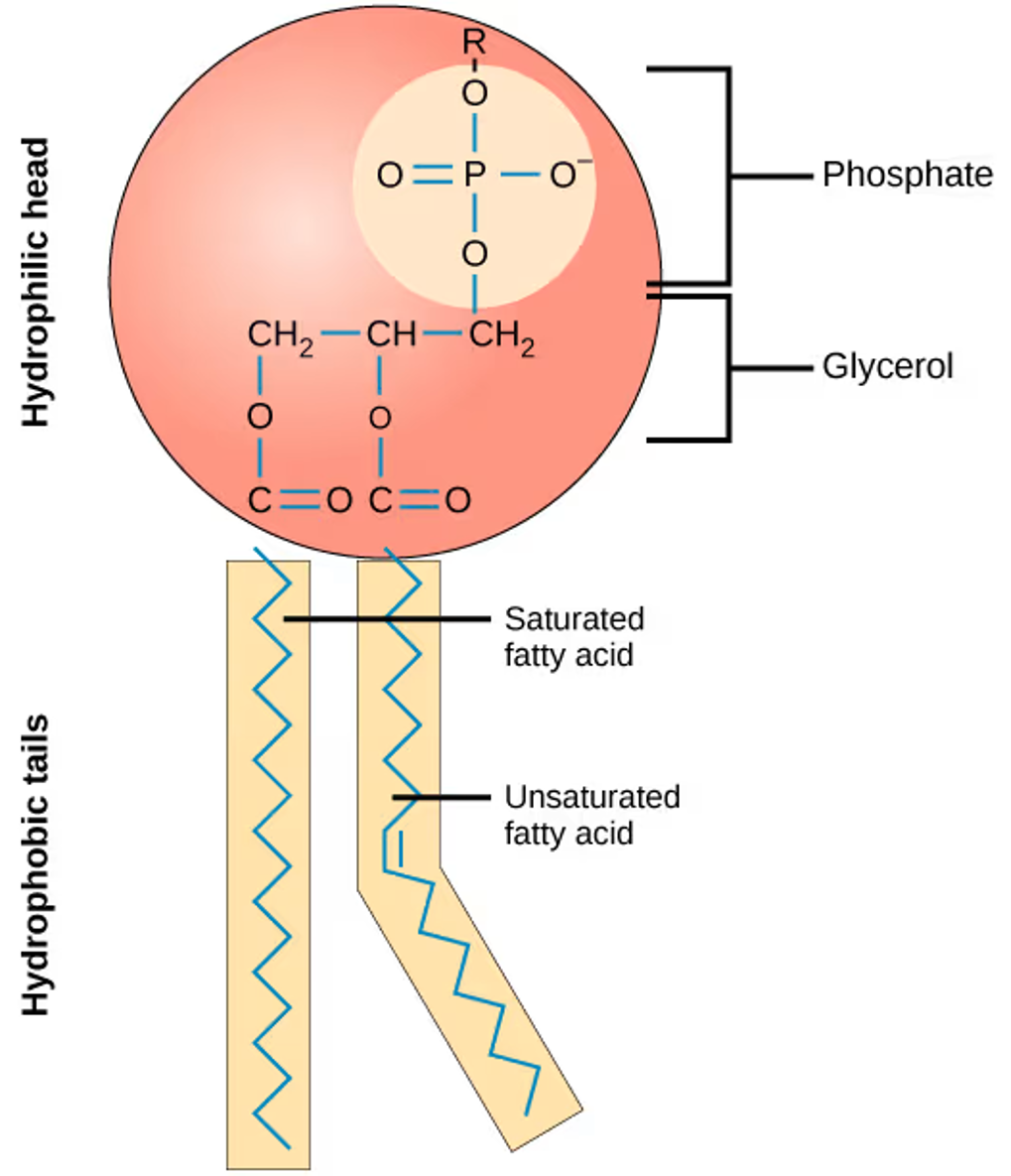
Phospholipids are helper lipids that contribute to the structural integrity and delivery performance of LNPs. Although they typically account for only 10–15 mol% of modern formulations, they play an important role in nanoparticle stability and membrane organization. The phospholipid DSPC is the most widely used and is present in several approved RNA-LNP therapeutics. Depending on their structure, phospholipids can influence both nanoparticle stability and intracellular delivery, making their selection an important parameter during LNP formulation development.
Sterol lipids
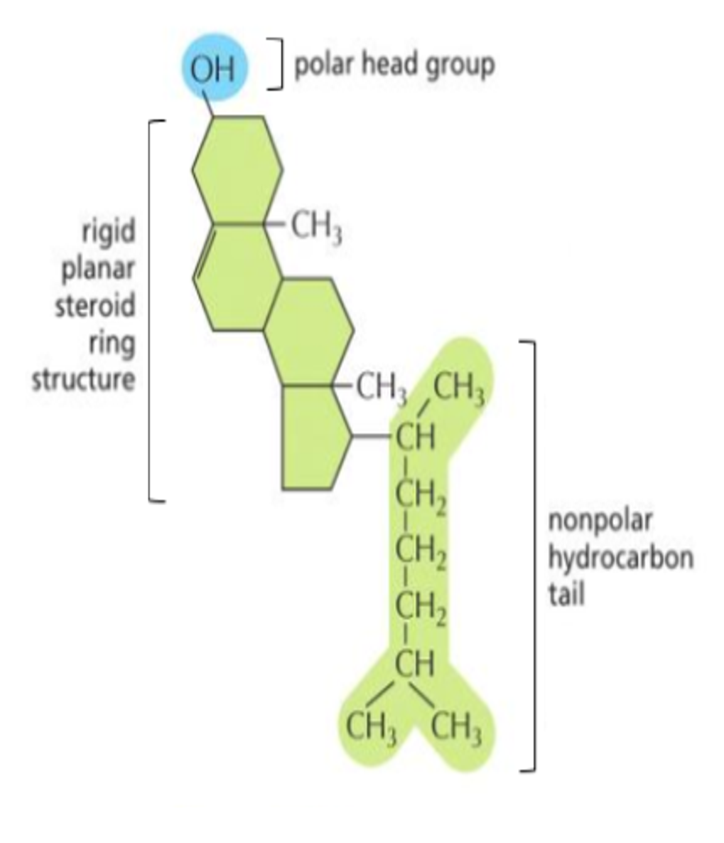
The sterol lipid – generally cholesterol – is an essential helper lipid that typically represents 30–50 mol% of an LNP formulation. By inserting between neighboring lipids, it modulates membrane organization, improves nanoparticle stability, and reduces premature cargo leakage. Cholesterol also contributes to membrane fusion processes and can influence the interaction of LNPs with biological membranes. As a result, cholesterol plays a key role in maintaining nanoparticle integrity while supporting the delivery of nucleic acid payloads.
Optimizing lipid composition is one of the most important aspects of RNA-LNP development, as changes in lipid types and molar ratios can significantly influence nanoparticle properties and biological performance. Table 2 summarizes the lipid compositions and molar ratios of clinically validated RNA-LNP formulations, including the Pfizer-BioNTech and Moderna COVID-19 mRNA-LNP vaccines.
Table 2. Lipid composition and molar ratios of representative clinically validated RNA-LNP formulations: Onpattro®, (Alnylam Pharmaceuticals), Comirnaty® (Pfizer-BioNTech), and Spikevax® (Moderna).
| Product name (Company) | Onpattro® (Alnylam) | Comirnaty® (Pfizer-BioNTech) | Spikevax® (Moderna) |
| Ionizable lipid | DLin-MC3-DMA (50%) | ALC-0315 (46.3%) | SM-102 (50%) |
| Phospholipid | DSPC (10%) | DSPC (9.4%) | DSPC (10%) |
| Sterol | Cholesterol (38.5%) | Cholesterol (42.7%) | Cholesterol (38.5%) |
| PEG-lipid | PEG2000-c-DMG (1.5%) | ALC-0159 (1.6%) | PEG2000-DMG (1.5%) |
| RNA modality | siRNA | mRNA | mRNA |
The lipid compositions summarized above also provide useful starting points for formulation development. Our LNP Starter Kits available in collaboration with CordenPharma can be used to evaluate established lipid combinations across RNA-LNP screening studies.
When preparing RNA-LNP formulations from individual lipid components, whether following an established composition or developing a custom formulation, molar ratios must be translated into practical preparation quantities. Our RNA-LNP Formulation Calculator helps determine the required lipid and RNA quantities for your formulation.
How are lipid nanoparticles synthesized and which method should you choose?
Why the LNP formulation process matters for drug development?
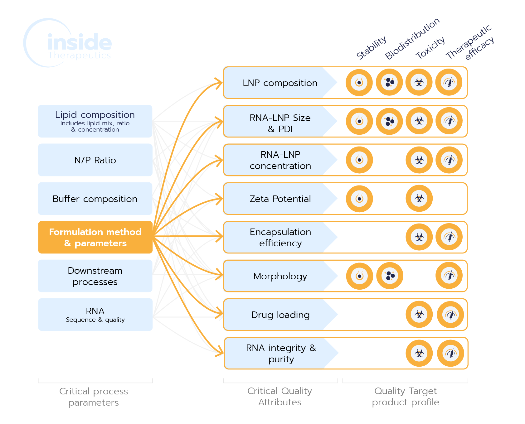
The LNP formulation process is pivotal in shaping Critical Quality Attributes (CQAs) like size, encapsulation efficiency, and morphology, which directly impact a drug’s efficacy, safety, and scalability. Choosing the right method ensures consistent, high-quality nanoparticles, reducing risks of formulation issues and facilitating smooth scale-up for clinical success.
Choice of LNP formulation method
A wide variety of methods are available for the synthesis of LNPs, many of which exploit solvent exchange to induce nanoprecipitation and lipid self-assembly in the presence of the nucleic acid cargo. Each approach offers a different trade-off in terms of particle characteristics, reproducibility, and scale-up capabilities. Selecting the appropriate formulation approach is therefore essential for successful RNA-LNP development, from early-stage screening to clinical manufacturing.
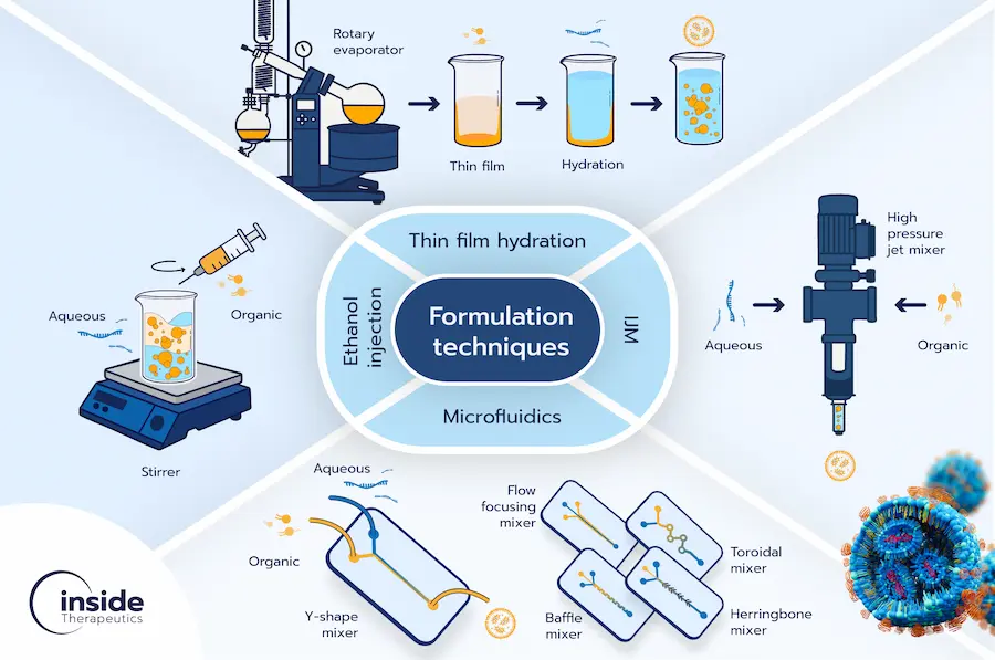
The current LNP manufacturing landscape ranges from simple manual and bulk preparation techniques to highly controlled macrofluidic and microfluidic platforms (Figure 8). Each approach presents distinct advantages and limitations depending on the stage of development and production requirements. A more detailed overview of these manufacturing technologies can be found in our LNP manufacturing review.
Manual methods
Manual approaches, such as hand mixing with pipette or in batch, rely on the spontaneous self-assembly of lipids and nucleic acids following the mixing of an organic lipid solution with an aqueous phase containing the cargo. Due to their simplicity and easy access, these methods are often used for preliminary proof-of-concept experiments.
While manual approaches can be employed for basic proof-of-concept experiments, they suffer from significant limitations. The lack of precise control over mixing conditions results in minimal control over final nanoparticle characteristics. This lack of consistency makes manual methods highly variable, unreliable, and unsuitable for reproducible or scalable nanoparticle production.
Traditional bulk methods: thin film hydration and ethanol injection
Traditional bulk methods, including thin film hydration and ethanol injection, remain widely used in academic research due to their accessibility and ease of implementation.
Thin film hydration involves dissolving lipids in an organic solvent, followed by solvent evaporation to form a thin lipid film. Upon hydration with an aqueous phase, lipids spontaneously self-assemble into vesicular structures. While straightforward to perform, this approach typically generates large and heterogeneous particles that often require additional processing steps such as extrusion to reduce size. Despite its simplicity, this method suffers from limited scalability, low encapsulation efficiency, and poor batch-to-batch reproducibility.
Ethanol injection follows a similar self-assembly principle, where a lipid-containing ethanol solution is rapidly injected into a stirred aqueous phase containing the nucleic acid cargo. Although simple and relatively reproducible at small scale, this approach also suffers from limited process control, batch-to-batch variability, low encapsulation efficiency, and poor scalability.
Macrofluidic approaches
Macrofluidic systems, such as T-mixers and impingement jet mixers (IJMs), provide significantly greater control over nanoparticle formation than conventional bulk methods. These technologies are highly effective for large-scale production.
In particular, impingement jet mixers (IJMs) generate rapid and homogeneous mixing by driving opposing fluid streams into a confined mixing chamber, supporting the formation of highly consistent nanoparticles. This technology offers a clear path toward industrial manufacturing through continuous processing and high-throughput production. IJMs played an important role in the manufacturing of mRNA-LNP vaccines during the COVID-19 pandemic.
Despite their advantages at manufacturing scale, IJMs operate within relatively constrained process conditions. Efficient mixing typically requires high total flow rates (TFRs), making particle formation highly sensitive to changes in flow conditions and resulting in relatively large minimum working volumes. In addition, the flow rate ratio (FRR) is generally constrained close to 1:1, limiting flexibility in formulation optimization. These requirements make IJMs less suitable for early-stage screening and preclinical workflows, where material availability is often limited and broader exploration of formulation parameters is required.
Microfluidic approaches
Microfluidic technologies have become the gold standard for RNA-LNP formulation development due to their exceptional control over nanoparticle self-assembly. In these systems, LNPs form through solvent exchange at the interface between an ethanol stream containing lipids and an aqueous stream containing nucleic acids.
The microscale dimensions enable rapid and highly reproducible mixing, resulting in homogeneous nanoparticle populations with tight control over size, polydispersity, and encapsulation efficiency. Importantly, process parameters such as flow rate ratio (FRR) and total flow rate (TFR) can be precisely adjusted to tune LNP properties according to the intended application. For a more detailed discussion of the underlying mixing mechanisms and micromixer architectures, see our review on microfluidics synthesis methods.
In addition to their high reproducibility, microfluidic platforms require only small sample volumes, making them particularly well suited for formulation screening, process optimization, and preclinical development. Typical RNA-LNP formulations produced by microfluidics achieve particle sizes between 50 – 200 nm with low polydispersity (PDI <0.2) and encapsulation efficiencies often exceeding 90%.
If you are evaluating microfluidic formulation for your RNA-LNP project — whether for initial lipid screening, formulation optimization, or the preparation of in vitro and in vivo study batches — see how TAMARA performs across a range of mRNA-LNP and siRNA-LNP formulations, including size, PDI, and encapsulation efficiency data across multiple lipid compositions.
Bridging LNP development and manufacturing scales
Moving from formulation development to manufacturing often requires switching mixing technologies, which can alter the conditions governing LNP formation and require additional process optimization. NanoPulse (Inside Therapeutics) was designed to address this process continuity challenge by enabling the same mixing principle to be maintained across a broad range of formulation volumes.
Unlike conventional microfluidic or IJM-based approaches, NanoPulse uses high-frequency alternating injection of the aqueous and lipid phases into a common channel. Mixing occurs at the interfaces generated between successive fluid segments, allowing formulation volume to be increased without changing the underlying mixing mechanism or being restricted to the specific process parameters.

Purification of RNA-LNP formulations
Regardless of the manufacturing approach selected, a downstream purification step is typically required to remove residual solvents and unencapsulated materials, thereby improving formulation stability and reducing the risk of toxicity prior to biological evaluation or clinical use.
Common purification strategies include dialysis, ultrafiltration, and tangential flow filtration (TFF). While dialysis and ultrafiltration is frequently used at laboratory scale due to their simplicity, TFF is generally employed for larger-scale applications because it enables efficient solvent exchange, concentration, and purification of LNP formulations with improved process control and scalability.
How are lipid nanoparticles characterized and what critical quality attributes matter?
The therapeutic performance of RNA-LNPs is highly dependent on their physicochemical properties. As discussed throughout this guide, even subtle variations in nanoparticle characteristics can influence stability, biodistribution, cellular uptake, and ultimately delivery efficiency.
Although a fully standardized regulatory framework for nanomedicines is still evolving, several characterization guidelines and best practices are available to support the development and evaluation of nanoparticle-based therapeutics. To ensure product quality and performance, a number of critical quality attributes (CQAs) must be carefully monitored.
Among the most important CQAs are particle size and size distribution (typically assessed through the polydispersity index, PDI), zeta potential, morphology, as well as nucleic acid loading characteristics such as encapsulation efficiency and drug loading. Together, these parameters provide valuable insights into nanoparticle quality and are often predictive of in vivo performance.
Size and size distribution
Particle size is one of the most important characteristics of RNA-LNPs, as it strongly influences their biological behavior in vivo. Size can affect circulation time, biodistribution, and cellular uptake, ultimately impacting therapeutic efficacy. LNP size is influenced by multiple formulation and process parameters, including lipid composition, formulation conditions, and manufacturing technology.
Therefore, the optimal LNP size varies depending on the target organ and application. While RNA-LNPs can be produced across a broad size range, most therapeutic formulations typically fall between 50 and 150 nm, balancing efficient delivery with favorable biodistribution profiles.
The polydispersity index (PDI) describes the width of the nanoparticle size distribution and provides an indication of population homogeneity. It is a dimensionless parameter defined as the square of the ratio between the standard deviation and the mean particle diameter (Eq. 1). PDI values range from 0 to 1, with lower values indicating more uniform nanoparticle populations.

In general, a PDI below 0.2 is considered highly homogeneous, while values below 0.3 are typically regarded as acceptable for RNA-LNP formulations across the industry. Microfluidic manufacturing technologies can routinely achieve highly homogeneous RNA-LNP populations with PDI values around 0.1 – 0.2, contributing to improved reproducibility and formulation consistency.

Interestingly, the size of lipid nanoparticles is primarily determined by the lipid composition and synthesis conditions rather than the size of the RNA cargo. As a result, formulation development often involves evaluating multiple lipid compositions and process conditions to identify formulations that achieve the desired physicochemical and biological properties. LNP starter kits can provide a convenient way to compare clinically relevant formulations during early-stage RNA-LNP development.
Particle size is commonly measured using techniques such as dynamic light scattering (DLS), nanoparticle tracking analysis (NTA), and laser diffraction (LD). The polydispersity index (PDI), however, is typically derived from DLS measurements.
Zeta potential
Zeta potential reflects the effective surface charge of nanoparticles in suspension. Rather than reflecting the true charge at the particle surface, it corresponds to the electrical potential at the so-called slipping plane, the boundary between the particle and the surrounding fluid as it moves through solution.
Zeta potential influences nanoparticle aggregation, protein adsorption, cellular uptake, and biodistribution, making it an important parameter to monitor during RNA-LNP development. Although near-neutral zeta potentials may increase the tendency for particle aggregation, the overall stability of RNA-LNPs also depends on steric stabilization mechanisms provided by other components such as PEG-lipids. On the other hand, charged nanoparticles may exhibit increased interactions with biological components, potentially affecting in vivo performance and toxicity. The optimal zeta potential depends on the intended application.
Zeta potential is most commonly measured using electrophoretic light scattering (ELS), where nanoparticle movement in an applied electric field is used to determine electrophoretic mobility and subsequently calculate the zeta potential.
Morphology and structure
RNA-LNPs do not possess a single, fixed internal structure. Multiple structural models have been proposed to describe RNA-LNP organization, reflecting the complexity of lipid–RNA interactions and the diversity of current formulations. Characterizing nanoparticle morphology and internal structure is therefore essential to better understand structure–function relationships in RNA delivery systems.
Cryogenic transmission electron microscopy (Cryo-TEM) is considered the gold standard for direct visualization of LNPs in their native hydrated state, allowing researchers to assess particle morphology, internal organization, and population heterogeneity. In addition to direct imaging methods, scattering techniques such as small-angle X-ray scattering (SAXS) and small-angle neutron scattering (SANS) provide information on the average internal structure of LNP populations. SAXS is commonly used to investigate features such as lipid organization, lamellarity, and layer thickness, while SANS can provide additional compositional information through contrast variation, enabling the localization of different nanoparticle components such as lipids and nucleic acids. Together, these techniques offer a comprehensive understanding of LNP morphology and structural architecture. A detailed overview of the major RNA-LNP structural models, including multilamellar, amorphous core, and bleb-containing morphologies, can be found in our comprehensive review on RNA-LNP morphology.

Encapsulation efficiency, encapsulation yield, and drug loading
Encapsulation efficiency (EE%), encapsulation yield (EY%), and RNA loading are key parameters used to evaluate the performance of RNA-LNP formulations. Together, they provide complementary information on formulation quality, process efficiency, and dosing capability.
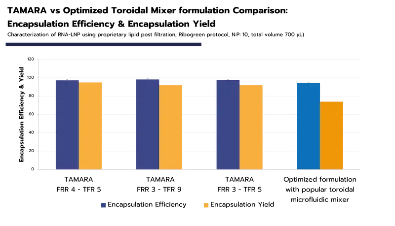
Encapsulation efficiency (EE%) represents the proportion of RNA encapsulated within the nanoparticles relative to the total RNA present in the sample (Eq. 2). It is one of the most widely reported quality attributes for RNA-LNP systems, as it directly impacts therapeutic potency and shows the formulation quality. Accurate determination of EE% is therefore essential during RNA-LNP development. For a step-by-step methodology, see our protocol on “Encapsulation Efficiency Analysis of RNA-LNPs Using the RiboGreen Assay“.

To facilitate data analysis, we also provide a downloadable RNA-LNP Encapsulation Efficiency (EE%) Calculator, available below.
Encapsulation yield (EY%) describes the overall recovery of encapsulated RNA relative to the initial RNA introduced during formulation (Eq. 3). Depending on when the measurement is performed, EY% can reflect losses occurring during nanoparticle formation alone or throughout both formulation and downstream processing. EY% becomes particularly important when working with limited or high-value RNA materials, where formulation losses can directly affect development timelines and costs. To explore real-world EE% and EY% data (Figure 11) generated with the TAMARA microfluidic platform in more details, including the impact of formulation parameters, production volume, and a direct comparison between TAMARA and toroidal mixer technologies, see our RNA-LNP formulation performance study.

RNA loading, sometimes referred to as drug loading (DL), quantifies the amount of RNA associated with the nanoparticles relative to the lipid content (Eq. 4). This parameter is particularly important for dosing considerations and is commonly expressed either as a weight percentage (wt%) or as micrograms of RNA per milligram of LNP.

Microfluidic-based RNA-LNP manufacturing technologies routinely achieve encapsulation efficiencies exceeding 90% for both mRNA and siRNA formulations. High EE%, EY%, and RNA loading values are generally desirable, as they contribute to efficient use of often costly RNA materials and support robust biological performance.
How do lipid nanoparticles deliver their payload inside target cells?
LNP delivery mechanism
Delivering nucleic acids to target cells is considerably more challenging than simply injecting them into the bloodstream. Once administered, free nucleic acids are rapidly exposed to multiple biological barriers. They are highly susceptible to degradation by endonucleases and can be cleared from circulation through uptake by cells of the immune system, particularly cells from the mononuclear phagocyte system (MPS). Depending on their size and physicochemical properties, some nucleic acid therapeutics may also undergo rapid renal clearance. Even if a fraction of the cargo survives all these barriers, these molecules still need to leave the bloodstream, penetrate target tissues, and cross the cellular membrane to reach its intracellular site of action.
RNA-LNPs have been developed to overcome these challenges by protecting nucleic acids from degradation, reducing their clearance, and facilitating cellular uptake and intracellular delivery. Surface modifications can further enhance biodistribution and enable selective targeting of specific tissues or cells.
The delivery process can be analyzed in three key steps: cellular uptake, intracellular trafficking, and endosomal escape. Successful completion of each step is essential for the RNA cargo to reach the cytoplasm and exert its biological activity.
Cellular uptake and intracellular delivery
Following intravenous administration, LNPs circulate throughout the body and and distribute to different tissues according to their physicochemical properties, including size, surface charge, composition, and surface functionalization.
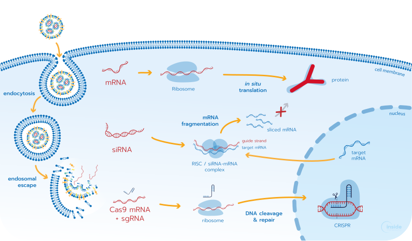
Following distribution into tissues, RNA-LNPs are primarily internalized by cells through endocytosis and trafficked through the endolysosomal pathway. During this process, endosomal vesicles progressively acidify as they mature from early endosomes (pH 6.0–6.5) to late endosomes (pH 5.0–5.5) and lysosomes (pH 4.5–5.0).
This acidic environment triggers the protonation of ionizable lipids, which become positively charged and interact with negatively charged endosomal membrane lipids. These interactions destabilize the endosomal membrane, promoting endosomal escape and releasing the nucleic acid cargo into the cytoplasm. Efficient endosomal escape is a critical determinant of RNA-LNP performance, as cargo that fails to escape is typically degraded within lysosomes or recycled back to the cell membrane. Because only a small proportion of internalized RNA successfully reaches the cytoplasm, endosomal escape is widely considered one of the major bottlenecks in RNA-LNP-mediated delivery.
Once released into the cytoplasm, the biological fate of the payload depends on its nature (Fig. 11). Messenger RNA (mRNA) is translated into proteins by ribosomes, while small interfering RNA (siRNA) associates with the RNA-induced silencing complex (RISC) to promote the degradation of complementary mRNA targets. In CRISPR-Cas9 applications, LNPs commonly co-deliver Cas9 mRNA and a single-guide RNA (sgRNA), enabling transient intracellular production of the Cas9 protein and subsequent gene editing.
To see an example of LNP-mediated CRISPR delivery in hematopoietic stem cells, explore our application note, “Benchmarking LNP vs Electroporation for eGFP RNA and CRISPR-Cas9 Delivery in HSCs“.
How can lipid nanoparticles be functionalized for targeted delivery?
Efficient intracellular delivery alone is not sufficient for a successful therapeutic. To maximize efficacy and minimize off-target effects, RNA-LNPs must also reach the appropriate tissue and cell population. Three main targeting strategies can be employed: passive, active, and endogenous targeting.
Passive targeting
Passive targeting relies on optimizing nanoparticle physicochemical properties — including size, surface charge, and hydrophilicity — to influence biodistribution and tissue accumulation.
One of the most widely studied passive targeting mechanisms is the Enhanced Permeability and Retention (EPR) effect observed in certain solid tumors. Due to their abnormal and leaky vasculature, tumor blood vessels may allow nanoparticles to extravasate more readily into the surrounding tissue. Combined with poor lymphatic drainage, this can promote local nanoparticle accumulation over time. Nanoparticle size plays an important role in this process, with particles typically in 20 – 200 nm range exhibiting favorable circulation and tissue penetration properties. PEGylation can further prolong circulation time by reducing clearance by the mononuclear phagocyte system (MPS). However, the magnitude of the EPR effect varies considerably between tumor types and experimental models.
Active targeting
Active targeting aims to improve cellular specificity through the incorporation of targeting ligands on the nanoparticle surface. These ligands recognize and bind specific receptors expressed on target cells, promoting preferential uptake of the therapeutic cargo.
A wide variety of targeting moieties can be employed, including antibodies, peptides, aptamers, carbohydrates, and small molecules. By exploiting receptor-ligand interactions, actively targeted LNPs can enhance delivery efficiency while reducing off-target exposure. However, achieving highly selective targeting remains challenging, as many receptors are also expressed on healthy tissues.
For a more detailed discussion of targeting strategies and ligand design, see our review on LNP targeting.
Endogenous targeting
Endogenous targeting leverages interactions between LNPs and naturally occurring plasma proteins that adsorb onto the nanoparticle surface after administration, forming a protein corona.
The composition of this protein corona is strongly influenced by the physicochemical properties of the LNP and can significantly affect biodistribution. A well-known example is the adsorption of apolipoprotein E (ApoE) onto LNPs, which promotes recognition by low-density lipoprotein receptors (LDLRs) expressed on hepatocytes and contributes to efficient liver targeting. This mechanism plays a key role in the clinical success of several approved RNA-LNP therapeutics.

Strategies for LNP surface functionalization
Several approaches can be used to incorporate targeting ligands onto the surface of RNA-LNPs.
In the pre-insertion approach, targeting ligands are first conjugated to lipid anchors, and incorporated into the lipid formulation prior to nanoparticle assembly. During LNP formation, these ligand-functionalized lipids become integrated into the nanoparticle structure. This strategy provides limited control over ligand orientation and surface accessibility, and sensitive ligands may be exposed to formulation conditions that can affect their integrity or activity.
In the post-insertion approach, ligand-functionalized lipids are prepared separately and introduced after nanoparticle formation. Upon mixing, these lipids insert into the outer membrane of preformed LNPs, increasing the likelihood that the targeting ligands remain exposed at the particle surface. Although this method requires an additional processing step, it offers greater control over ligand presentation.
Finally, post-formulation surface conjugation involves the direct attachment of targeting ligands onto preformed LNPs containing reactive surface groups. This approach enables selective surface functionalization through established bioconjugation chemistries. However, it generally requires additional synthesis and purification steps.

Lipid nanoparticle applications
Thanks to their ability to protect and efficiently deliver nucleic acids into cells, LNPs have emerged as versatile platform technology with applications across vaccines, gene therapy, oncology, and personalized medicine. By enabling cells to produce or silence specific proteins, or perform targeted genome editing, RNA-LNPs open new therapeutic possibilities for disease prevention and treatment.
mRNA vaccines
mRNA-LNPs have revolutionized vaccine development by enabling the rapid delivery of genetic instructions encoding a target antigen. Following cellular uptake, the delivered mRNA is translated into proteins that stimulate an immune response and promote the production of protective antibodies.
This approach gained worldwide recognition through the development of COVID-19 mRNA vaccines. Beyond infectious diseases, RNA-LNP vaccines are also being investigated for personalized cancer immunotherapy, where patient-specific tumor antigens can be used to stimulate anti-tumor immune responses.
Gene & cell therapies
Gene therapy aims to treat diseases by modifying, replacing, or regulating gene expression. RNA-LNPs provide a non-viral delivery platform for a wide range of genetic medicines, including mRNA, siRNA, micro RNA (miRNA), antisense oligonucleotides (ASOs), and CRISPR-Cas9 systems. Depending on the therapeutic strategy, these molecules can be used to produce missing proteins, silence disease-causing genes, or perform targeted genome editing. As a result, RNA-LNPs are being actively explored for the treatment of genetic disorders, rare diseases, and inherited conditions.
Additionally, RNA-LNP technologies are increasingly being explored for cell-engineering applications, including ex vivo and in vivo immune cell programming and emerging CAR-T–related strategies.
Drug delivery
Beyond nucleic acid therapeutics, LNPs can also be used to encapsulate and deliver conventional drugs. By enhancing solubility, protecting payloads from degradation, and improving biodistribution, they can increase the therapeutic effectiveness of drug candidates.
Particularly in oncology, LNP-based delivery systems are being investigated to increase drug accumulation within tumors while reducing systemic toxicity.
Personalized medicine
The programmable nature of RNA therapeutics makes RNA-LNPs particularly attractive for personalized medicine. By tailoring the RNA sequence to the genetic profile of an individual patient or disease, treatments can be adapted to specific mutations, biomarkers, or tumor antigens.
How do lipid nanoparticles compare to other nanoparticle delivery platforms?
LNPs are part of a broader ecosystem of nanoparticle delivery technologies developed for drug and nucleic acid delivery applications. While LNPs have become leading platforms for RNA therapeutics, several alternative systems continue to be explored.
Solid lipid nanoparticles (SLNs), developed in the 1990s, use a solid lipid matrix to improve the physical stability of encapsulated compounds and modulate drug release profiles. Although initially designed for hydrophobic small molecules, SLNs have also been investigated for nucleic acid delivery through the incorporation of cationic lipids or surfactants capable of interacting with negatively charged RNA or DNA molecules. However, due to their relatively dense and dehydrated lipid core, nucleic acids are often associated with the particle surface rather than efficiently entrapped within the nanoparticle structure, which can limit cargo protection, intracellular release, and overall transfection performance compared with ionizable lipid-based LNP systems. For a detailed overview of lipid-based platforms and their structural differences, see our review on lipid-based nanoparticles (LBNPs).
Polymer nanoparticles (PNPs), including systems based on polymers such as poly(lactic-co-glycolic acid) (PLGA), offer broad chemical versatility and tunable degradation kinetics. Several PLGA-based formulations have received regulatory approval for small-molecule drug delivery applications. In the context of nucleic acid therapeutics, polymeric systems are being actively investigated for applications requiring controlled release, enhanced stability, or alternative targeting strategies, although intracellular delivery performance can vary significantly depending on formulation design.
Peptide-based nanoparticles (PBNs) represent another emerging class of non-viral delivery systems built from synthetic or naturally derived peptides, including cell-penetrating peptides. Their adaptable chemistry and favorable biocompatibility profiles make them attractive candidates for targeted therapeutic applications. In addition, peptide engineering can enable receptor-specific interactions and improved cellular uptake, although susceptibility to proteolytic degradation remains an important consideration for in vivo applications.
While polymer- and peptide-based nanoparticles continue to show strong therapeutic potential and are rapidly evolving, LNPs currently represent the most clinically advanced and industrially established non-viral platform for nucleic acid delivery. Their widespread use in RNA therapeutics has accelerated the development of scalable manufacturing processes, regulatory familiarity, and clinical translation strategies.
Lipid nanoparticles: Key takeaways and future perspectives
LNPs have become a key platform for nucleic acid therapeutics, enabling RNAs to be protected, transported to cells, and released intracellularly to exert their biological function. Their performance depends on the careful design of the formulation and control of process parameters, which together determine LNP physicochemical properties — including particle size, PDI, RNA encapsulation, and morphology — and ultimately influence biodistribution, cellular uptake, endosomal escape, and therapeutic activity.
Developing effective RNA-LNPs therefore requires the systematic optimization of formulation composition and process conditions for the intended payload and application. Alongside optimization, comprehensive characterization is essential to understand how these parameters affect critical quality attributes and to establish reproducible formulations with the desired biological performance.
Despite significant progress in RNA-LNP development, several challenges continue to shape the future of the field. A deeper understanding of LNP internal structure and its relationship with biological performance is still needed, while inefficient endosomal escape remains a major limitation to intracellular RNA delivery. New alternatives to conventional PEG-lipids are also being explored to address concerns related to immune responses and repeated administration. In parallel, active targeting and lipid composition-based strategies are expanding opportunities for delivery beyond the liver. From a manufacturing perspective, the challenge is increasingly to maintain consistent process conditions and LNP quality as formulations transition from small-scale development to larger-scale production.
As RNA therapeutics continue to expand across vaccines, gene therapy, and oncology, advances in LNP composition, targeting, intracellular delivery, characterization, and scalable manufacturing will be central to translating these emerging modalities into effective therapies.
For practical guidance beyond this review, explore our RNA-LNP resources, including experimental protocols, formulation calculator, application notes, and technologies for RNA-LNP development and scale-up.
References
[1] R. Tenchov, J. M. Sasso, and Q. A. Zhou, “PEGylated Lipid Nanoparticle Formulations: Immunological Safety and Efficiency Perspective,” Bioconjug. Chem., vol. 34, no. 6, pp. 941–960, Jun. 2023, doi: 10.1021/ACS.BIOCONJCHEM.3C00174.
[2] N. Miralles, “Asymmetric Lipid Transfer Between Vesicles Measured by QCM-D,” 2021.
[3] C. J. Stadtherr, “Cholesterol, part 1: What is it?” Accessed: Aug. 06, 2026. [Online]. Available: https://revitalizemetabolichealth.com/cholesterol-and-its-role-in-health-pt-1/
[4] T. Bethiana et al., “Identifying differential effects from eleven mixing techniques on mRNA lipid nanoparticle physicochemistry and biological performance,” Nov. 09, 2025. doi: 10.1101/2025.11.07.687311.
[5] S. Gül, J. Vergnaud, F. Fay, and E. Fattal, “Precision engineering of macrophage reprogramming with RNA interference-loaded lipid nanoparticles: a game-changer in cancer immunotherapy,” Drug Deliv. Transl. Res., vol. 16, no. 7, pp. 2272–2301, Jul. 2026, doi: 10.1007/S13346-025-01970-1.
[6] I. Menon, M. Zaroudi, Y. Zhang, E. Aisenbrey, and L. Hui, “Fabrication of active targeting lipid nanoparticles: Challenges and perspectives,” Mater. Today Adv., vol. 16, p. 100299, Dec. 2022, doi: 10.1016/J.MTADV.2022.100299.

Looking to get started with RNA-LNP formulation?
Reach out to us to learn how we can help!
Frequently Asked Questions on Lipid Nanoparticles
What is a lipid nanoparticle (LNP)?
A lipid nanoparticle (LNP) is a nanoscale delivery vehicle designed to protect and transport therapeutic payloads, particularly nucleic acids such as mRNA and siRNA, into target cells. RNA-LNPs are composed of a mixture of lipids that self-assemble into nanoparticles with a formulation-dependent internal structure. They are the delivery platform behind the COVID-19 mRNA vaccines and currently represent the most clinically advanced non-viral system for nucleic acid therapeutics.
What’s the difference between an LNP and a liposome?
The key difference is the ionizable lipid. Conventional liposomes typically contain neutral or permanently charged lipids. Neutral lipids are generally less effective at encapsulating negatively charged RNA, whereas permanently cationic lipids improve RNA encapsulation but are associated with higher cytotoxicity and nonspecific interactions. In contrast, RNA-LNPs incorporate ionizable lipids that become positively charged under acidic conditions during formulation to promote efficient RNA encapsulation, remain largely neutral at physiological pH to improve tolerability, and are protonated again within endosomes to facilitate intracellular RNA release.
What is a lipid nanoparticle typically made of?
A standard RNA-LNP formulation combines four lipid components: an ionizable lipid (40–50 mol%), a helper phospholipid such as DSPC (10–15 mol%), cholesterol (30–50 mol%), and a PEG-lipid (1.5–2 mol%). Each component plays a distinct role — the ionizable lipid drives encapsulation and endosomal escape, cholesterol stabilizes the particle, and PEG-lipids control circulation time and colloidal stability.
Why is microfluidics considered the best method for early-stage LNP formulation?
Microfluidics is considered the gold standard for early-stage LNP formulation because it gives precise, reproducible control over nanoparticle self-assembly at low sample volumes. By tuning flow rate ratio (FRR) and total flow rate (TFR), microfluidic systems routinely produce LNPs with PDI below 0.20 and encapsulation efficiency above 90% — making them well suited for screening and preclinical development, where manual mixing and bulk methods fall short on consistency and scalability.
What is the main bottleneck in LNP-mediated RNA delivery?
Endosomal escape is widely considered the biggest bottleneck in LNP-mediated RNA delivery. After cellular uptake, only a small fraction of the RNA cargo escapes the endosome before it’s degraded in lysosomes or recycled back to the cell surface — making endosomal escape efficiency a critical determinant of how much of the delivered RNA actually reaches the cytoplasm to act.