
Contact
Any inquiries about your nanoparticle/RNA-LNP formulation?
Drop us a line!
Loading specific message...

Contact
Any inquiries about your nanoparticle/RNA-LNP formulation?
Drop us a line!
Loading specific message...
Book a demo
Tell us about your nanoparticle or RNA-LNP project and request a TAMARA demonstration.
Our team will get back to you shortly to schedule your session.
Loading specific message...
Home Resources Application notes
In vivo evaluation of mRNA-LNP vaccines formulated using TAMARA
Abstract
mRNA-based vaccines have emerged as a powerful platform for the prevention of infectious diseases, enabling rapid antigen design and in situ protein expression. Their success critically depends on efficient delivery systems capable of ensuring robust expression and immune activation in vivo. Lipid nanoparticles (LNPs) have become the leading non-viral platform for RNA delivery, protecting mRNA from degradation while facilitating cellular uptake and intracellular release.
Here, we evaluate the in vivo performance of mRNA-LNPs formulated using the TAMARA microfluidic system. ALC-0315-based LNP formulations were administered intramuscularly in mice, and their performance was assessed through analysis of antigen expression, biodistribution, and immune responses. Bioluminescence imaging revealed strong and sustained expression at the injection site, with transient systemic distribution. Immunological analyses demonstrated robust antigen-specific antibody responses, significantly enhanced following booster immunization, as well as strong antigen-specific cellular responses.
Together, these results demonstrate that mRNA-LNPs formulated using TAMARA enable efficient in vivo delivery and potent immune activation, supporting their relevance as a platform for vaccine development.
Evaluation of antigen expression, biodistribution, and immune responses of mRNA-LNP vaccines in vivo.
Introduction
The emergence of mRNA-based vaccines has transformed approaches to the prevention of infectious diseases. By enabling rapid design and transient in situ expression of antigenic proteins, mRNA technologies provide a highly adaptable platform capable of responding to both emerging and established pathogens. However, the effectiveness of these approaches relies on the ability to deliver mRNA efficiently into target tissues while ensuring sufficient protein expression to trigger a protective immune response.
Lipid nanoparticles (LNPs) serve as the primary non-viral platform for RNA delivery, providing protection against degradation and promoting efficient entry into cells followed by intracellular release. Their clinical validation in mRNA COVID-19 vaccines has demonstrated their capacity to support efficient in vivo delivery. Importantly, their modular composition allows precise tuning of physicochemical properties and biological performance, enabling optimization of biodistribution, expression kinetics, and immune activation.
During the development of mRNA-LNP vaccines, a comprehensive evaluation of in vivo performance remains essential. Key parameters include the level and duration of antigen expression, the extent of systemic distribution, and the ability to induce both humoral and cellular immune responses. Together, these factors determine the overall efficacy and safety profile of vaccine candidates.
In this study, mRNA-LNPs formulated using the TAMARA microfluidic system were evaluated following intramuscular (IM) administration in mice. The in vivo performance of ALC-0315-based formulations was assessed through the analysis of antigen expression, biodistribution, and immune responses. This work provides an integrated evaluation of mRNA-LNP vaccine performance, linking delivery efficiency to downstream immunological outcomes.
This study was conducted in collaboration with the HUN-REN Biological Research Centre in Szeged, with contributions from Csaba Bajusz, and supported by Inside Therapeutics.
Results
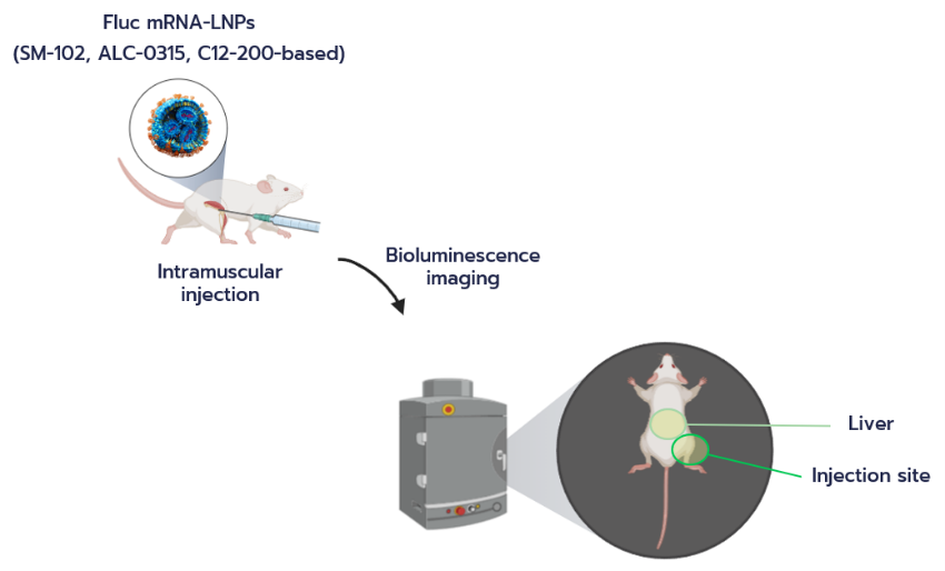
1/ In vivo expression and biodistribution of mRNA-LNPs
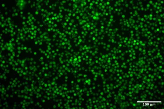
Following IM administration of luciferase (Luc)-encoding mRNA-LNPs in BALB/c mice, in vivo expression and biodistribution were monitored over time using bioluminescence imaging (Fig. 1). Both wild-type luciferase and a tagged luciferase construct were evaluated to assess the impact of payload design on expression dynamics.

At the injection site, strong bioluminescent signals were observed at early time points for both constructs, indicating efficient local delivery and translation of the mRNA cargo (Fig. 2). Signal intensity gradually decreased over time, consistent with the transient nature of mRNA expression. While the tagged construct exhibited slightly lower signal intensity compared to the wild-type luciferase, it remained robustly expressed throughout the duration of the study.
In the liver, detectable bioluminescent signals were observed at early time points for both constructs, reflecting partial systemic distribution of the nanoparticles following intramuscular injection (Fig. 2). However, in contrast to the injection site, the liver signal declined rapidly over time and reached low baseline levels within a few days. This faster decay indicates transient systemic exposure and limited persistence of off-target expression.

Overall, these results demonstrate efficient and sustained antigen expression at the injection site, supporting the suitability of these mRNA-LNP formulations for in vivo applications.
2/ Humoral immune response of mRNA-LNPs
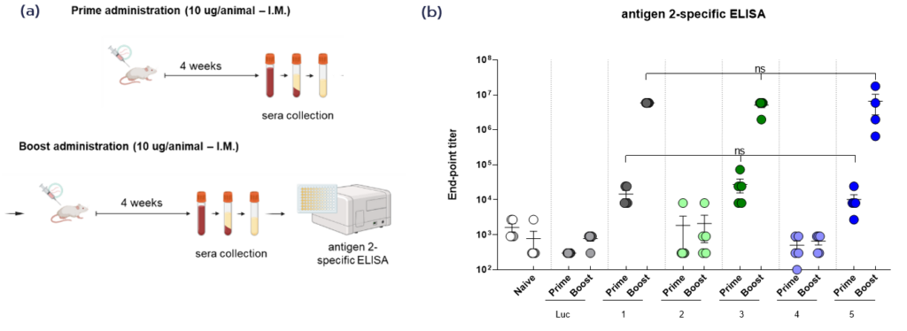
The ability of mRNA-LNP formulations to induce antigen-specific antibody responses was evaluated using a prime–boost immunization strategy. Mice received IM administrations of mRNA-LNPs encoding different antigen constructs, and serum samples were collected four weeks after the primary injection and four weeks following the booster dose to assess the magnitude of the humoral response (Fig. 3a).
Antigen-specific antibody titers were measured by ELISA, revealing marked differences between the tested constructs (Fig. 3b). Following the primary immunization, moderate antibody responses were detected for selected constructs (1, 3, and 5), indicating successful antigen expression and initial immune priming. In contrast, other constructs (2 and 4) induced minimal or negligible responses.
Administration of a booster dose led to a substantial increase in antibody titers for the most effective formulations (Fig. 3b). In particular, constructs 1, 3, and 5 elicited strong antigen-specific responses, with titers increasing by several orders of magnitude compared to the primary response. This pronounced boost effect reflects effective immune memory formation and amplification of the humoral response. Conversely, constructs 2 and 4 remained weakly immunogenic, showing no improvement after boosting.
Control groups, including naïve animals and luciferase mRNA-LNP-treated mice, exhibited minimal antibody responses across all conditions, confirming the specificity of the observed immune responses (Fig. 3b). Overall, these results demonstrate that mRNA-LNP delivery enables robust antigen-specific antibody production in vivo, with a clear dependence on construct design and a strong enhancement upon booster immunization.
3/ Cellular immune response of mRNA-LNPs
To evaluate the ability of mRNA-LNP formulations to elicit antigen-specific cellular immune responses, ELISpot assays were performed on splenocytes isolated from mice 12 days after IM administration. Cells were stimulated ex vivo under three conditions — no stimulus, ConA stimulation, and antigen-specific stimulation — to assess baseline activity, overall immune competence, and antigen-specific cellular immune responses, respectively (Fig. 4a).
Under unstimulated conditions, all groups exhibited minimal spot formation, indicating low baseline cytokine production and confirming the absence of nonspecific activation (Fig. 4b). Upon stimulation with ConA, a potent mitogenic activator used as a positive control, all groups displayed strong responses, demonstrating that the isolated immune cells remained viable and functionally competent.
In contrast, antigen-specific stimulation revealed a clear and selective response in mice immunized with antigen-encoding mRNA-LNPs (Fig. 4b). A substantial increase in spot numbers was observed in the antigen 1 group compared to both naïve and luciferase control groups, which remained at baseline levels. This indicates that the cellular immune response was specifically induced by the antigen encoded in the mRNA-LNP formulation, rather than by nonspecific immune activation.
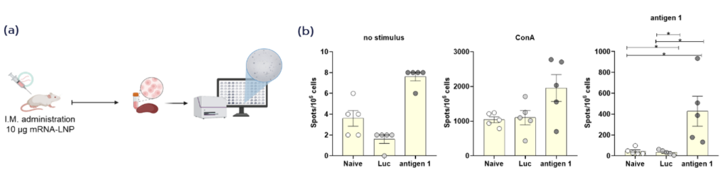
Overall, these results demonstrate that mRNA-LNP delivery effectively induces antigen-specific cellular immune responses in vivo, complementing the humoral responses observed and confirming the ability of these formulations to trigger a comprehensive adaptive immune response.
Conclusion & discussions
This study provides a comprehensive evaluation of the in vivo performance of mRNA-LNP formulations following intramuscular administration. Efficient antigen expression was achieved at the injection site, with sustained signal over time and transient systemic distribution. These delivery characteristics are critical for ensuring sufficient antigen exposure while minimizing off-target effects.
The immunological analyses further demonstrate the ability of mRNA-LNPs to induce a robust adaptive immune response. Strong antigen-specific antibody responses were observed for selected constructs, with a marked increase following booster immunization, indicating effective immune priming and memory formation. In parallel, antigen-specific cellular responses were detected, confirming activation of immune pathways essential for vaccine efficacy.
Importantly, variability observed between mRNA constructs highlights the influence of antigen design on immunogenicity, emphasizing the need for systematic screening and optimization during vaccine development. The combination of efficient delivery and tunable formulation properties enables mRNA-LNP platforms to accommodate such optimization strategies.
Taken together, these results demonstrate that mRNA-LNPs formulated using TAMARA support efficient in vivo delivery and the induction of both humoral and cellular immune responses.
Materials & methods
1/ mRNA-LNP formulation using TAMARA
mRNA-loaded lipid nanoparticles (LNPs) were formulated using the TAMARA microfluidic system (Inside Therapeutics). An ALC-0315-based lipid composition adapted from clinically validated formulations was used, consisting of ALC-0315/DSPC/cholesterol/DMG-PEG at a molar ratio of 50/10/38.5/1.5. Lipids were dissolved in ethanol at a total lipid concentration of 30 mM. mRNA constructs encoding luciferase or antigen sequences were encapsulated during the formulation process.
LNPs were produced by controlled mixing of an ethanolic lipid phase with an aqueous mRNA solution prepared in 25 mM sodium acetate buffer (pH 4.0, mRNA concentration: 0.314 µg/µL) under defined flow conditions (FRR=3, TFR= 2 mL/min). Following formulation, nanoparticles were purified by dialysis to remove residual solvent and exchange buffer.
Further details on the formulation procedure can be found in the “RNA-LNP Formulation Protocol” available on the Inside Therapeutics website.
2/ Characterization
The hydrodynamic diameter and polydispersity index (PDI) of the mRNA-LNPs were determined by dynamic light scattering (DLS) using a Litesizer DLS 100 instrument (Anton Paar, Austria). The resulting LNPs exhibited sizes around 90 nm, with low PDI values indicating homogeneous nanoparticle populations (Luc mRNA-LNP: 0.15; tagged-Luc mRNA-LNP: 0.13; other mRNA-LNP constructs: 0.14–0.20).
RNA concentrations and encapsulation efficiencies (EE%) were determined using Qubit RNA High Sensitivity (Thermo Fisher Scientific, Q32852) and Broad Range (Thermo Fisher Scientific, Q10210) Assay Kits with a Qubit 4 Fluorometer (Thermo Fisher Scientific). All formulations exhibited encapsulation efficiencies above 98%, while unencapsulated mRNA levels remained below the detection limit of the assay kits.
3/ Animal studies
All in vivo experiments were performed using six- to eight-week-old inbred female wild-type BALB/cOlaHsd mice (Envigo). mRNA-LNP formulations were administered via intramuscular injection into the musculus gastrocnemius at the indicated doses.
For biodistribution and expression studies, mice received 5 µg of luciferase-encoding mRNA-LNPs. For immunogenicity studies, mice were immunized with 10 µg of mRNA-LNPs encoding antigen constructs. In a prime–boost regimen, a booster dose was administered four weeks after the primary injection.
4/ In vivo bioluminescence imaging
In vivo expression of luciferase was assessed using an In Vivo Imaging System (IVIS) Lumina III imaging system (PerkinElmer). Following intramuscular administration of luciferase mRNA-LNPs, mice were injected intraperitoneally with D-luciferin (150 mg/kg). Bioluminescent signals were measured at multiple time points post-injection.
Signal intensity was quantified as total flux (photons per second) in regions of interest corresponding to the injection site and liver.
5/ ELISA for humoral immune response
Antigen-specific antibody responses were evaluated by enzyme-linked immunosorbent assay (ELISA). Serum samples were collected four weeks after the primary immunization and four weeks following the booster dose.
Antibody titers were determined by measuring antigen-specific binding and expressed as endpoint titers.
6/ ELISpot for cellular immune response
Cellular immune responses were assessed using Enzyme-Linked ImmunoSpot assays (ELISpot). Splenocytes were isolated from mice 12 days after intramuscular administration of mRNA-LNPs.
Cells were stimulated ex vivo under three conditions: no stimulation, Concanavalin A (ConA) stimulation as a positive control, and antigen-specific stimulation. The number of cytokine-secreting cells was quantified as spot-forming units per number of plated cells using the Mabtech IRIS system.
Acknowledgements
Figures 1, 3a, and 4a were created using BioRender.com.


Looking to get started or improve your LNP formulation screening?
Reach out to us to discover how we can help!
Other Application notes
Looking for more detailed application notes on nanoparticle formulations? Check out our other resources!
See all Application notes