
Contact
Any inquiries about your nanoparticle/RNA-LNP formulation?
Drop us a line!
Loading specific message...

Contact
Any inquiries about your nanoparticle/RNA-LNP formulation?
Drop us a line!
Loading specific message...
Book a demo
Tell us about your nanoparticle or RNA-LNP project and request a TAMARA demonstration.
Our team will get back to you shortly to schedule your session.
Loading specific message...
Home Resources Application notes
In vivo evaluation of standard mRNA-LNP formulations produced with TAMARA
Abstract
RNA-based therapeutics offer a versatile platform for disease intervention, supported by rapid design and precise programmability. Their clinical implementation, however, is limited by susceptibility to degradation and inefficient cellular delivery. Lipid nanoparticles (LNPs) have emerged as the primary delivery system, enabling RNA protection and intracellular transport, with demonstrated clinical success in siRNA therapeutics and mRNA and saRNA vaccines.
In this study, mRNA-LNPs produced using the TAMARA microfluidic system were evaluated across three standard lipid formulations: SM-102, ALC-0315, and C12-200. Following intramuscular administration in mice, in vivo expression and biodistribution were assessed using firefly luciferase (Fluc) as a reporter.
All formulations enabled efficient in vivo expression, with formulation-dependent differences in signal intensity at the injection site (SM-102 > ALC-0315 > C12-200). Expression was transient and predominantly localized at the injection site after an initial phase of systemic distribution.
Comparative analysis of in vivo expression across clinically relevant lipid nanoparticle systems.
Introduction
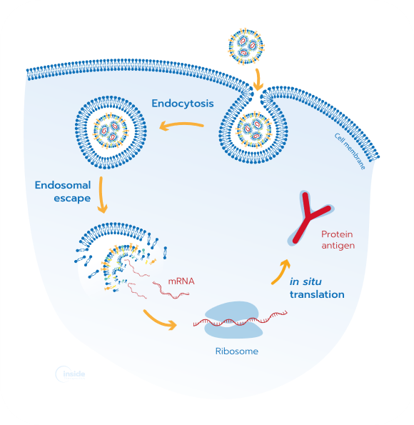
RNA-based therapeutics have emerged as a powerful and adaptable platform for disease treatment and prevention, driven by their rapid design, sequence programmability, and ability to enable transient protein expression. These features allow for fast response to emerging medical needs and precise control over therapeutic function. However, the successful application of RNA in vivo remains constrained by its inherent instability and susceptibility to rapid degradation, as well as the need for efficient delivery into target cells.
Lipid nanoparticles (LNPs) have become the leading solution to these challenges, providing protection of RNA cargo while facilitating cellular uptake and intracellular release. Their modular composition enables fine-tuning of physicochemical and biological properties, allowing optimization for specific applications. The clinical success of LNP-based therapeutics, including siRNA drugs and mRNA and saRNA vaccines, has established their central role in enabling RNA delivery at scale and in vivo functionality.
The performance of LNPs is highly dependent on formulation parameters, including lipid composition and manufacturing conditions, which influence particle size, encapsulation efficiency, morphology, and ultimately in vivo behavior. Microfluidic technologies have emerged as powerful tools for LNP formulation, enabling controlled and reproducible mixing conditions that are critical for consistent nanoparticle production. In this context, the TAMARA microfluidic platform offers a robust approach for the formulation of mRNA-LNPs under well-defined and scalable conditions, from early screening to in vivo studies.
Here, mRNA-LNPs were produced using TAMARA across three widely used lipid systems — SM-102, ALC-0315, and C12-200 — and evaluated for their physicochemical properties and in vivo performance. Using firefly luciferase (Fluc) mRNA as a reporter, expression and biodistribution were assessed following intramuscular (IM) administration in mice, enabling comparison of formulation-dependent differences in vivo.
This work was carried out at the University of Strathclyde in the laboratory of Prof. Yvonne Perrie by MSc Jade Forrester, and supported by PhD Claire Counil, PhD Audrey Nsamela and PhD Sezen Gul from Inside Therapeutics.
Results
1/ Physicochemical characterization of mRNA-LNPs
mRNA-LNPs were formulated using the TAMARA microfluidic system with three widely used ionizable lipid compositions: SM-102, ALC-0315, and C12-200. To assess the impact of process parameters on nanoparticle properties, LNPs were produced at two total flow rates (TFR 1 and 5 mL/min), and their size and polydispersity index (PDI) were measured in triplicate using dynamic light scattering (DLS).
Across all lipid formulations, increasing the total flow rate from 1 to 5 mL/min resulted in a reduction in particle size, indicating effective control of nanoparticle size through mixing conditions (Fig. 1). This decrease in size was accompanied by a modest increase in PDI; however, values remained within an acceptable range, reflecting the formation of homogeneous particle populations.

Importantly, the effect of TFR on both size and PDI was consistent across all lipid systems, demonstrating the robustness and reproducibility of the TAMARA platform independently of lipid composition.
The formulations produced at TFR 5 mL/min were selected for in vivo studies. Under this condition, all LNPs exhibited high encapsulation efficiency (EE%), exceeding 90% as measured by the RiboGreen assay, indicating efficient RNA loading across lipid systems.
Overall, these results demonstrate that TAMARA enables the reproducible production of mRNA-LNPs with tunable size and high encapsulation efficiency across standard lipid formulations, providing a solid foundation for subsequent in vivo evaluation.
2/ In vivo expression & biodistribution of mRNA-LNPs
The in vivo performance of TAMARA-formulated mRNA-LNPs was evaluated following IM administration in mice using Fluc mRNA as a reporter. Bioluminescence imaging was performed across multiple time points to monitor expression, and quantitative analysis of total flux was used to assess signal intensity at both the injection site and in the liver. Imaging was conducted in both dorsal and ventral views, with ventral imaging providing higher signal intensity and improved sensitivity for analysis; therefore, only ventral images are presented in this application note.
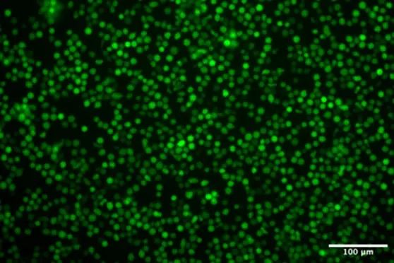
All formulations enabled detectable in vivo expression, confirming successful delivery and functional translation of the mRNA cargo (Fig. 2). Strong bioluminescent signals were observed at early time points (Day 0–2) across all lipid systems, indicating rapid onset of expression following administration (Fig. 2a). Signal intensity decreased progressively over time, becoming undetectable in the representative images after approximately one week due to image scaling and reduced intensity. This behavior is consistent with the transient nature of mRNA expression in vivo.
At early time points, signal intensity was higher in the liver compared to the injection site, suggesting initial systemic distribution of a fraction of the administered LNPs (Fig. 2b). Over time, liver-associated signal declined more rapidly, while expression at the injection site persisted longer.
Quantitative total flux analysis further highlighted formulation-dependent differences in expression levels. Across all time points at the injection site, SM-102-based LNPs exhibited the highest signal intensity, followed by ALC-0315 and C12-200. While the overall expression kinetics were similar across formulations, characterized by rapid onset and gradual decay, the magnitude of expression differed depending on lipid composition.
Together, these results demonstrate that TAMARA-formulated mRNA-LNPs enable efficient and transient in vivo expression. The observed formulation-dependent differences further highlight the utility of TAMARA as a platform for comparative screening and optimization of LNPs.

Conclusion & discussions
The results demonstrate that the TAMARA microfluidic platform enables the reproducible formulation of mRNA-LNPs with controlled physicochemical properties, including tunable particle size and high encapsulation efficiency, supporting consistent in vivo performance.
All formulations showed efficient mRNA expression following intramuscular administration, with rapid onset and transient signal consistent with the expected kinetics of mRNA-based systems. The progressive decay in signal reflects the non-integrating nature of mRNA and its physiological degradation in vivo. Furthermore, differences in signal intensity across formulations highlight the critical role of lipid composition in determining in vivo performance, with SM-102 showing the highest expression levels.
Overall, TAMARA enables reliable production and comparison of mRNA-LNPs, supporting efficient formulation screening and optimization for RNA-based therapeutics.
Materials & methods
1/ mRNA-LNP formulation using TAMARA
Lipid nanoparticles (LNPs) were formulated using the TAMARA microfluidic platform (Inside Therapeutics) with three ionizable lipid compositions: SM-102, ALC-0315, and C12-200 (Table 1). Firefly luciferase (Fluc) mRNA, provided by CPI (Centre for Process Innovation, Sedgefield, UK), was used as cargo. Lipids were dissolved in ethanol as the organic phase. For SM-102 and ALC-0315 formulations, the aqueous phase consisted of citrate buffer (50 mM, pH 5.0), whereas C12-200 formulations were prepared using sodium acetate/sodium chloride buffer (50 mM sodium acetate, 100 mM sodium chloride, pH 4.0). Microfluidic mixing was performed at a flow rate ratio (FRR) of 3:1 and total flow rates (TFR) of 1 or 5 mL/min, with an N/P ratio of 6. Nanoparticles were purified by ultrafiltration using Amicon centrifugal filters, with Tris buffer (10 mM, pH 5.0) used as the purification buffer.
Table 1. Lipid composition and molar ratios of mRNA-LNP formulations.
| SM-102 | ALC-0315 | LP-01 | |
| Ionizable lipid | SM-102 (50%) | ALC-0315 (46.3%) | LP-01 (45%) |
| Phospholipid | DSPC (10%) | DSPC (9.4%) | DSPC (9%) |
| Sterol | Cholesterol (38.5%) | Cholesterol (42.7%) | Cholesterol (44%) |
| PEG-lipid | MPEG-2000-DMG (1.5%) | ALC-0159 (1.6%) | MPEG-2000-DMG (2%) |
Further details on the formulation procedure can be found in the “RNA-LNP Formulation Protocol” available on the Inside Therapeutics website.
2/ Physicochemical characterization
Particle size (Z-average) and polydispersity index (PDI) were measured by dynamic light scattering (DLS). Measurements were performed in triplicate. Encapsulation efficiency (EE%) was determined using a RiboGreen assay.
3/ In vivo studies
mRNA-LNPs were administered intramuscularly into the hind limbs of BALB/c mice at a dose of 2 µg mRNA per animal (1 µg per leg). Bioluminescence imaging was performed following subcutaneous injection of D-luciferin, and images were acquired at multiple time points post-administration. Both dorsal and ventral images were collected, with ventral images used for analysis in this application note.
4/ Bioluminescence imaging
Bioluminescent signal was quantified as total flux (photons per second, p/s) using region-of-interest (ROI) analysis at the injection site and in the liver. Data were collected over time to assess expression kinetics and signal distribution.
Acknowledgements
We acknowledge CPI for supplying the Fluc mRNA used in the study, which was produced under the Intracellular Drug Delivery Centre program, funded by Innovate UK (project number: 10058505).

Looking to get started or improve your LNP formulation screening?
Reach out to us to discover how we can help!
Other Application notes
Looking for more detailed application notes on nanoparticle formulations? Check out our other resources!
See all Application notes