


Book a demo
Tell us about your nanoparticle or RNA project and request Ntensify® micro and mini demo
Our team will get back to you shortly to schedule your session.
Loading specific message...
Contact
Any inquiries about RNA synthesis systems?
Drop us a line!
Loading specific message...

Contact
Any inquiries about your nanoparticle/RNA-LNP formulation?
Drop us a line!
Loading specific message...

Contact
Any inquiries about your nanoparticle/RNA-LNP formulation?
Drop us a line!
Loading specific message...
Book a demo
Tell us about your nanoparticle or RNA-LNP project and request a TAMARA demonstration.
Our team will get back to you shortly to schedule your session.
Loading specific message...
Home Resources Application notes
Integrating High-Purity mRNA Synthesis and Scalable LNP Formulation: AMCAP™ and TAMARA for RNA-LNP Development
Abstract
The successful clinical translation of messenger RNA (mRNA) therapeutics critically depends on achieving high-quality mRNA constructs and developing safe, efficient delivery systems. This application note details a synergistic strategy that combines Anemocyte’s innovative AMCAP™ one-pot enzymatic capping system for high-purity, 5’ CAP-1 mRNA synthesis with Inside Therapeutics’ high-precision TAMARA microfluidic platform for scalable lipid nanoparticle (LNP) formulation. This integrated workflow significantly simplifies manufacturing by reducing time, cost, and process complexity compared to conventional methods. Formulation of two AMCAP™ mRNA constructs of different lengths into LNPs using TAMARA yielded particles with controlled size, high stability (confirmed by DLS), and high in vitro transfection efficiency.
This validated, integrated approach offers a reproducible and scalable solution for mRNA-LNP optimization, supporting high-quality mRNA production, robust LNP formulation, and advanced RNA therapeutic applications.
From one-pot mRNA synthesis to microfluidic LNP formulation.
Introduction
In recent years, messenger RNA (mRNA) has emerged as a promising new therapeutic agent to prevent and treat various diseases. The delivery of mRNA offers a versatile platform for achieving rapid and robust protein expression.
However, its intrinsic lability makes robust delivery systems crucial for protection against degradation, efficient cellular uptake, and release into the cytosol. The successful clinical translation of mRNA relies on two interconnected pillars:
- The synthesis of high-quality, pure constructs;
- The development of safe, efficient delivery systems.
The Delivery Mechanism: Lipid Nanoparticles (LNPs)
Lipid nanoparticles (LNPs) have become the leading delivery platform for nucleic acids, as evidenced by the rapid development of LNP-based mRNA vaccines (e.g., COVID-19) (1, 2), which highlighted their scalability, safety, and therapeutic efficacy. Beyond vaccines, LNPs are now actively being explored for applications including gene editing, protein replacement therapies, and cancer immunotherapy.
LNPs are the preferred delivery system due to their intrinsic ability to protect fragile nucleic acids and facilitate efficient cellular uptake, while enabling cytoplasmic delivery through endosomal escape (3). Their physicochemical properties (e.g., size distribution, surface charge) are readily adaptable, and their modular lipid composition allows for customization for distinct therapeutic objectives.
LNPs are typically composed of four distinct lipid components combined at specific molar ratios to facilitate nucleic acid encapsulation and delivery (4,5):
- Ionizable cationic lipids (40 – 50 mol%) play the primary role in encapsulating and delivering nucleic acids. At low pH during formulation, they enable efficient electrostatic complexation with RNA, driving nanoparticle self-assembly. Post-uptake, they become protonated in the acidic endosomal environment, facilitating RNA release by destabilizing endosomal membranes (6–8). Examples include SM-102 (Moderna), ALC-0315 (Pfizer-BioNTech), and LP-01 (Novartis).
- Cholesterol (38 – 50 mol%) is a critical structural component that enhances stability, modulates membrane fluidity and integrity, and contributes to endocytosis. In recent years, other cholesterol derivatives, such as β-sitosterol, have also been explored to further tune LNP properties (8).
- Polyethylene glycol (PEG)-lipids (1.5 – 2.0 mol%) form a protective hydrophilic layer on LNPs, which increases their stability and circulation time, reduces recognition by the immune system and helps control particle size and distribution (7). Commonly used PEG-lipids include PEG-DMG (Moderna), and ALC-0159 (Pfizer-BioNTech) (9).
- Helper phospholipids (10.0 – 15.0 mol%) are essential in LNP formulation to support structural integrity and modulate the fusogenicity of LNPs. DSPC, DOPE, and DOPC are frequently used. Notably, DSPC is used in two marketed COVID-19 vaccines BNT162b2 and mRNA-1273 (10).
Anemocyte’s Innovative mRNA Manufacturing Platform
High-fidelity mRNA synthesis necessitates stringent control over the capping process, a modification indispensable for molecular stability and high translational efficiency.
Anemocyte has addressed this challenge through the development of a novel enzymatic ONE-POT reaction capping system (AMCAP™), that allows fast and efficient synthesis of 5’ CAP-1 mRNA in a single step. This innovative system employs T7 RNA polymerase in conjunction with the Vaccinia capping system, enabling a unified synthesis and capping reaction.
Compared to the standard two steps and the co-transcriptional capping reactions, Anemocyte process significantly simplifies the manufacturing workflow, in terms of time, costs and processing steps, while maintaining high mRNA quality (Figure 1).

Addressing Manufacturing Challenges with TAMARA Microfluidic Platform
Despite significant technological advancements, LNP manufacturing remains challenging, as different formulation approaches introduce trade-offs between accessibility, reproducibility, process control, and scalability. Achieving efficient and highly reproducible LNP synthesis is therefore essential for successful RNA-LNP development and clinical translation.
Microfluidic production has emerged as a key platform for preclinical RNA-LNP development, enabling highly controlled synthesis that yields excellent particle size uniformity and high encapsulation efficiency.
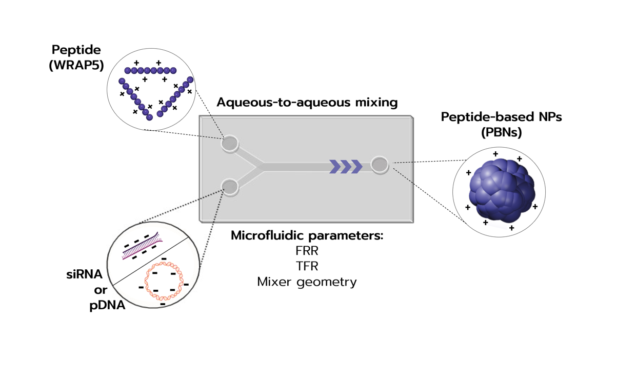
Inside Therapeutics offers a robust solution to these manufacturing hurdles: the TAMARA nanoparticle formulation platform (Figure 2). TAMARA is a validated, plug-and-play microfluidic system designed for reproducible and scalable LNP manufacturing, supporting applications from early-stage formulation screening to preclinical studies.

Figure 2. Workflow of LNP formulation using the TAMARA microfluidic system.
Anemocyte and Inside Therapeutics: A Synergistic Approach to mRNA-LNP Optimization
Anemocyte and Inside Therapeutics combine complementary expertise across the mRNA-LNP value chain, spanning mRNA synthesis and high-precision formulation. Anemocyte’s strategic focus encompasses proprietary mRNA production and optimized construct design, complemented by advanced microfluidic technologies from Inside Therapeutics for controlled and reproducible LNP formulation. Together, these complementary technologies enable the development of high-quality, well-defined mRNA-LNP systems.
This application note demonstrates the benefits of integrating AMCAP™ and TAMARA into a streamlined workflow for reproducible RNA-LNP development.
Experimental workflow:
As shown in Figure 3, the streamlined workflow consists of the following steps:
- ONE-POT synthesis and purification of AMCAP™ mRNA;
- Preparation of individual lipid stock solutions;
- Preparation of AMCAP™ mRNA solution (aqueous phase) and lipid mix (organic phase);
- Setting of microfluidic process parameters;
- Microfluidic mixing using the TAMARA platform;
- LNP-encapsulated mRNA (mRNA-LNP) formulation;
- Physical characterization and biological validation of mRNA-LNPs.

Results
1/ DLS analysis of mRNA-LNPs
mRNA1-LNP and mRNA2-LNP formulations were prepared using the TAMARA microfluidic platform with the SM-102 LNP kit at a fixed nitrogen-to-phosphate (N/P) ratio of 6 and a final volume of 1 mL. The starting concentration for both mRNA constructs was 115 µg/mL, with a 12.5 mM lipid mixture. Following formulation, each sample was dialysed overnight at room temperature. DLS was performed before and after dialysis (3 runs/sample, backscattering detection), reporting the Z-average size and polydispersity index (PDI).
Table 1. Formulation parameters and DLS characterization of mRNA1-LNPs before and after dialysis.
| Sample name | Flow rate ratio (FRR) | Total flow rate (TFR; mL/min) | Pre-dialysis size (nm) | Pre-dialysis PDI | Post-dialysis size (nm) | Post-dialysis PDI |
| mRNA1A-LNPs | 3 | 1 | 128.3 | 0.08 | 121.9 | 0.06 |
| mRNA1B-LNPs | 3 | 5 | 81.3 | 0.17 | 92.3 | 0.15 |
| mRNA1C-LNPs | 3 | 10 | 61.1 | 0.25 | 85.5 | 0.17 |
| mRNA1D-LNPs | 4 | 5 | 80.2 | 0.15 | 115.4 | 0.06 |
| mRNA1E-LNPs | 2 | 5 | 96.0 | 0.11 | 89.6 | 0.08 |
Table 2. Formulation parameters and DLS characterization of mRNA2-LNPs before and after dialysis.
| Sample name | Flow rate ratio (FRR) | Total flow rate (TFR; mL/min) | Pre-dialysis size (nm) | Pre-dialysis PDI | Post-dialysis size (nm) | Post-dialysis PDI |
| mRNA2A-LNPs | 3 | 1 | 133.8 | 0.06 | 128.9 | 0.06 |
| mRNA2B-LNPs | 3 | 5 | 84.6 | 0.11 | 97.6 | 0.15 |
| mRNA2C-LNPs | 3 | 10 | 70.0 | 0.20 | 101.4 | 0.12 |
| mRNA2D-LNPs | 4 | 5 | 80.9 | 0.11 | 90.6 | 0.11 |
| mRNA2E-LNPs | 2 | 5 | 88.6 | 0.10 | 101.0 | 0.08 |
At a fixed FRR of 3, increasing the TFR from 1 to 10 mL/min resulted in a decrease in particle size for both mRNA1-LNPs and mRNA2-LNPs, while maintaining low PDI values, indicative of uniform particle populations.
The effect of FRR on particle size was less pronounced compared to TFR. At a constant TFR of 5 mL/min, increasing the FRR from 2 to 4 led to a modest reduction in particle size for mRNA2-LNPs pre- and post-dialysis, and for mRNA1-LNPs pre-dialysis, while a slight increase was observed for mRNA1-LNPs post-dialysis. Overall, PDIs remained low across all formulations, indicating particle uniformity.
According to the Inside Therapeutics LNP Starter Kit Guide, SM-102-based LNPs formulated under these conditions typically achieve encapsulation efficiencies above 80%, corresponding to an estimated encapsulated mRNA concentration of approximately 90 µg/mL.
2/ Delivery of mRNA-LNPs
2.1/ Expression of EGFP mRNA-LNPs
To evaluate Enhanced Green Fluorescent Protein (EGFP) mRNA expression in vitro, HEK293T cells were treated with different doses of lipid nanoparticle (LNP)-encapsulated mRNA1 (mRNA1-LNPs), ranging from 0.25 µg/well up to 2.00 µg/well, in IMDM medium supplemented with 2% FBS according to standard procedures as described in the Materials & Methods section.
As a positive control, the cells were transfected with 1 μg or 0.5 μg of naked EGFP mRNA from Anemocyte (AMCAP™ EGFP mRNA) using JetMessenger (Polyplus). After 72 hours of incubation at +37 °C and 5% CO2, cells were observed by fluorescence microscopy (Figure 4A) and then analyzed by flow cytometry using a Sony SH 800 (Sony Biotechnology) (Figure 4B and Figure 5).

As presented in Figure 4A and 4B, HEK293T cells transfected with five different mRNA1-LNPs (using AMCAPTM EGFP mRNA formulated with the SM-102-based lipids mix) showed high transfection efficiency, with a percentage of EGFP positive cells greater than 90%. This result was comparable to the positive control (HEK293T cells transfected with 1 μg of AMCAPTM mRNA1).
Overall, all tested mRNA1-LNP formulations exhibited robust EGFP expression, which was comparable to, or slightly higher than the expression level observed with the mRNA1 transfected using JetMessenger. Specifically, mRNA1A-LNPsandmRNA1B-LNPs, which were prepared at a fixed Flow Rate Ratio (FRR) of 3 and Total Flow Rates (TFRs) of 1 and 5 mL/min showed the highest expression levels at the maximum dose tested (Figure 5).
Furthermore, mRNA1D-LNPs and mRNA1E-LNPs prepared at a constant TFR of 5 mL/min with FRRs of 4 and 2, respectively, sustained strong EGFP expression, even when the dose was reduced by half. Across all formulations, EGFP expression increased in a dose-dependent manner (Figure 5).

of mRNA1-LNPs. Data reported as Mean of Fluorescence Intensity (MFI).
2.2/ Expression of β-GAL mRNA-LNPs
To evaluate βeta Galactosidase (β-GAL) enzymatic activity, HEK293T cells were seeded in a 96-well plate at a cell density of 12,500 cells/well in 0.2 mL of IMDM (Thermo Fisher Scientific) supplemented with 10% FBS (Thermo Fisher Scientific). After 24 hours of incubation at +37 °C and 5% CO2, the cells were treated with different amounts of mRNA2-LNPs, ranging from 0.4 µg/well up to 1.7 µg/well, in IMDM supplemented with 2% FBS.
As a positive control, the cells were transfected with 0.2 μg, 0.1 μg or 0.05 μg of naked βeta galactosidase (β-GAL) mRNA from Anemocyte (AMCAPTM β-GAL mRNA) following standard procedures with JetMessenger (Polyplus). After 72 hours, cells were analyzed using the luminescence-based BETA-GLO assay system (Promega) (Figure 5). Data is reported as a fold induction of activity compared to untreated cells.

All the mRNA2-LNP (β-GAL) formulations induced a high β-galactosidase expression. The enzymatic activity was consistently high, with fold change values ranging from 14.2 up to 15.0 across the tested doses (1.7 to 0.4 µg/well). These expressions levels were comparable to the maximum dose used for the β-GAL mRNA transfected using JetMessenger (0.2 µg, fold change 14.2), indicating efficient delivery and subsequent β-galactosidase expression and activity.
Conclusion & discussions
This collaborative study highlights the complementary strengths of high-purity mRNA synthesis using AMCAP™ and reproducible RNA-LNP formulation using TAMARA.
The AMCAP™ ONE-POT capping system enabled rapid production of high-quality 5′ CAP-1 mRNA while simplifying the manufacturing workflow by reducing processing time, costs, and workflow complexity. The TAMARA microfluidic platform generated reproducible mRNA-LNP formulations with controlled particle size and low PDI across different process conditions, demonstrating its suitability for robust and scalable RNA-LNP production.
The formulated mRNA-LNPs exhibited efficient in vitro delivery and high protein expression for both EGFP and β-gal mRNA constructs, confirming the compatibility of the integrated workflow from mRNA synthesis through LNP formulation.
Together, AMCAP™ and TAMARA provide a streamlined approach to mRNA-LNP development, supporting efficient manufacturing, formulation optimization, and the development of high-performance RNA therapeutics.
Materials & methods
1/ IVT reaction
The AMCAP™ mRNA-LNP formulation using the TAMARA microfluidic system by Inside Therapeutics provides a simple, fast, and scalable solution for producing a variety of mRNA-LNP formulations suitable for a broad range of therapeutic applications. The mRNA used for lipid combinations must be of a high quality, pure construct to ensure that the resulting formulation enables safe and efficient delivery.
The In vitro Transcription (IVT) reaction was performed starting from linearized plasmids encoding for Enhanced Green Fluorescent Protein (EGFP) mRNA or βeta galactosidase (β-GAL) mRNA. The transcribed mRNA sequences for both EGFP and β-Gal contain a 5’ untranslated (UTR) region, a 3’ UTR sequence and a polyA tail. In vitro transcription was performed utilizing the AMCAP™ ONE-POT synthesis and capping method. The mRNA obtained was then purified using affinity chromatography. Concentration and buffer exchange were carried out using Tangential Flow Filtration (TFF). Concentrated mRNA was resuspended in sodium citrate and stored at -80°C prior to the formulation step.
2/ mRNA-LNP formulation and characterization
mRNA-LNP formulations were prepared using the TAMARA microfluidic platform in a staggered herringbone mixer (SHM) configuration. The SM-102 LNP starter kit (CordenPharma) was used for all formulations. Lipids were dissolved in ethanol to obtain the organic phase at an initial concentration of 12.5 mM, while mRNAs were diluted to an initial concentration of 115 µg/mL in an aqueous buffer (10.0 mM citrate buffer, pH 4.0).
Formulations were generated by controlled microfluidic mixing of the aqueous and organic phases under defined flow rate ratios (FRR) and total flow rates (TFR), as specified in Table 1 and Table 2. All formulations were prepared at a fixed nitrogen-to-phosphate (N/P) ratio of 6 and produced at a final volume of 1 mL.
Following microfluidic mixing, LNPs were purified by dialysis. Particle size and polydispersity index (PDI) were measured by dynamic light scattering (DLS) both before and after purification.
Further details on the formulation procedure can be found in the “RNA-LNP Formulation Protocol” available on the Inside Therapeutics website.
3/ Cell transfection
HEK293 cells were seeded 24 hours before transfection in 24-well or 96-well plates at a cell density of 70,000 or 12,500 cells/well, respectively, in 0.5 mL or 0.2 mL of Iscove’s Modified Dulbecco’s Medium (IMDM) (Thermo Fisher Scientific) supplemented with 10% Foetal Bovine Serum (FBS) (Thermo Fisher Scientific).
After 24 hours of incubation at +37 °C and 5% CO2, the cells were treated with different amounts of mRNA1-LNPs (EGFP) or mRNA2-LNPs (β-GAL) in IMDM medium supplemented with 2% FBS. As positive control, cells were transfected with naked mRNA1 or mRNA2 at different concentrations using JetMessenger (Polyplus) following the manufacturer’s instructions.
Transfected cells were incubated for 72 hours at +37 °C and 5% CO2 and then analyzed by flow cytometry using Sony SH 800 (Sony Biotechnology) for EGFP expression or by enzymatic assay (Beta-GLO, Promega) for βeta -Galactosidase mRNA.


Looking to get started or improve your LNP formulation screening?
Reach out to us to discover how we can help!
Other Application notes
Looking for more detailed application notes on nanoparticle formulations? Check out our other resources!
See all Application notes
From microfluidic formulation to payload quantification: TAMARA and CloudSpec for RNA-LNP development
Read more
In vivo evaluation of standard mRNA-LNP formulations produced with TAMARA
Read more
In vivo evaluation of mRNA-LNP vaccines formulated using TAMARA
Read more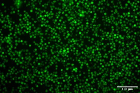
Benchmarking LNP vs Electroporation for eGFP RNA and CRISPR-Cas9 Delivery in HSCs
Read more
Streamlining mRNA-LNP screening with TAMARA
Read more
